

Search Count: 33
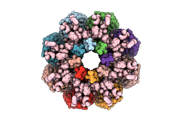 |
Structure Of Sticholisin Ii In Large Unilamellar Vesicles.
Organism: Stichodactyla helianthus
Method: ELECTRON MICROSCOPY Release Date: 2025-10-29 Classification: MEMBRANE PROTEIN Ligands: FO4, CLR |
Organism: Stichodactyla helianthus
Method: ELECTRON MICROSCOPY
Release Date: 2025-10-29
Ligands: FO4, CLR
 |
Structure Of Fragacetoxin C In Lipid Nanodiscs
Organism: Actinia fragacea
Method: ELECTRON MICROSCOPY Release Date: 2025-10-29 Classification: MEMBRANE PROTEIN Ligands: FO4, CLR |
Organism: Actinia fragacea
Method: ELECTRON MICROSCOPY
Release Date: 2025-10-29
Ligands: FO4, CLR
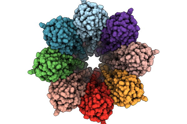 |
Structure Of The Octameric Pore Of Fragaceotxin C (Frac Or Delta-Actitoxin-Afr1A) In Large Unilamellar Vesicles.
Organism: Actinia fragacea
Method: ELECTRON MICROSCOPY Release Date: 2025-10-08 Classification: MEMBRANE PROTEIN Ligands: FO4, CLR |
Organism: Actinia fragacea
Method: ELECTRON MICROSCOPY
Release Date: 2025-10-08
Ligands: FO4, CLR
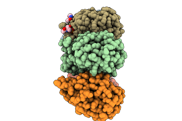 |
Structure Of 6Mer Pore Intermediate Of Sticholysin Ii (Stnii) Toxin In Lipid Nanodiscs
Organism: Stichodactyla helianthus
Method: ELECTRON MICROSCOPY Release Date: 2025-10-08 Classification: MEMBRANE PROTEIN Ligands: FO4 |
Organism: Stichodactyla helianthus
Method: ELECTRON MICROSCOPY
Release Date: 2025-10-08
Ligands: FO4
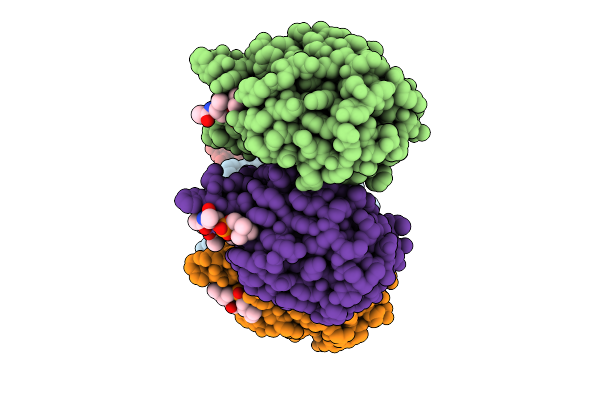 |
Structure Of 5Mer Pore Intermediate Of Sticholysin Ii (Stnii) Toxin In Lipid Nanodiscs
Organism: Stichodactyla helianthus
Method: ELECTRON MICROSCOPY Release Date: 2025-09-17 Classification: MEMBRANE PROTEIN Ligands: FO4 |
Organism: Stichodactyla helianthus
Method: ELECTRON MICROSCOPY
Release Date: 2025-09-17
Ligands: FO4
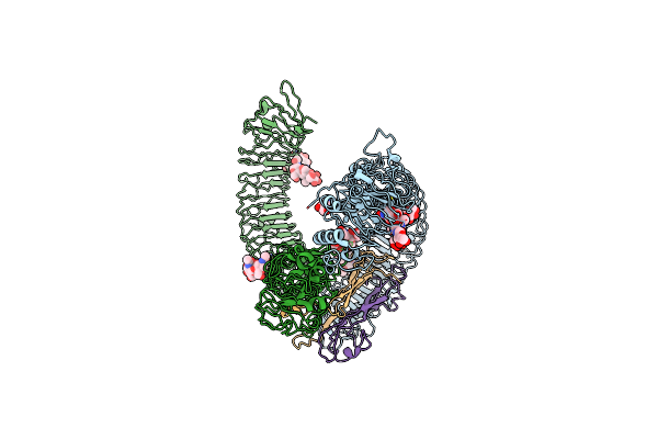 |
Cryo-Em Of Aedes Aegypti Toll5A Dimer Bound To Spz1C
Organism: Aedes aegypti
Method: ELECTRON MICROSCOPY Release Date: 2022-07-13 Classification: IMMUNE SYSTEM Ligands: NAG |
Organism: Aedes aegypti
Method: ELECTRON MICROSCOPY
Release Date: 2022-07-13
Ligands: NAG
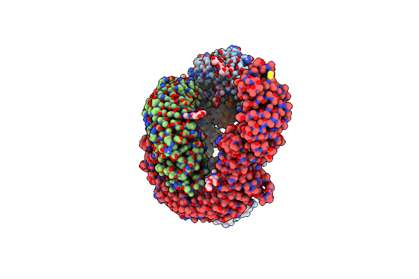 |
Cryo-Em Of Aedes Aegypti Toll5A Trimer Bound To Spz1C
Organism: Aedes aegypti
Method: ELECTRON MICROSCOPY Release Date: 2022-07-13 Classification: IMMUNE SYSTEM Ligands: NAG |
Organism: Aedes aegypti
Method: ELECTRON MICROSCOPY
Release Date: 2022-07-13
Ligands: NAG
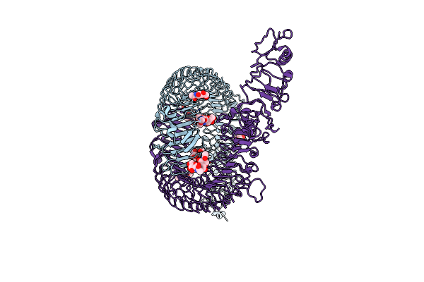 |
Cryo-Em Of Aedes Aegypti Toll5A
Organism: Aedes aegypti
Method: ELECTRON MICROSCOPY Release Date: 2022-07-13 Classification: IMMUNE SYSTEM Ligands: NAG |
Organism: Aedes aegypti
Method: ELECTRON MICROSCOPY
Release Date: 2022-07-13
Ligands: NAG
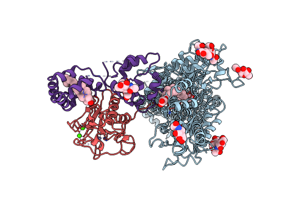 |
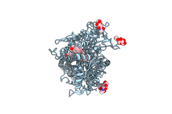
Cryo-Em Structure Of Xenopus Patched-1 In Complex With Gas1 And Sonic Hedgehog
Organism: Xenopus laevis, Homo sapiens
Method: ELECTRON MICROSCOPY Release Date: 2022-03-02 Classification: MEMBRANE PROTEIN Ligands: CLR, Y01, NAG, ZN, CA, PLM |
Organism: Xenopus laevis, Homo sapiens
Method: ELECTRON MICROSCOPY
Release Date: 2022-03-02
Ligands: CLR, Y01, NAG, ZN, CA, PLM
 |
Cryo-Em Structure Of Xenopus Patched-1 In Nanodisc
Organism: Xenopus calcaratus
Method: ELECTRON MICROSCOPY Release Date: 2022-03-02 Classification: MEMBRANE PROTEIN Ligands: CLR, NAG |
Organism: Xenopus calcaratus
Method: ELECTRON MICROSCOPY
Release Date: 2022-03-02
Ligands: CLR, NAG
 |
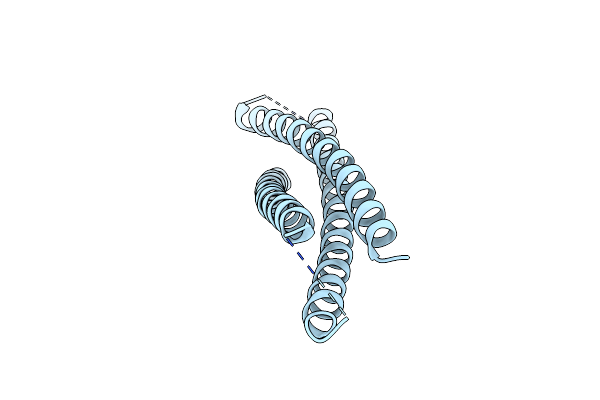
X-Ray Structure Of The Apo Form Of The Establishement Gene Regulator Reg576 Of The G+ Plasmid P576
Organism: Bacillus altitudinis
Method: X-RAY DIFFRACTION Resolution:1.98 Å Release Date: 2018-10-17 Classification: TRANSCRIPTION |
Organism: Bacillus altitudinis
Method: X-RAY DIFFRACTION
Release Date: 2018-10-17
 |
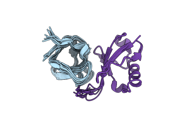
Crystal Structure Of A Rationally Designed Single-Chain Protein Mimicking A Trimeric Gp41 N-Terminal Heptad-Repeat Region
Organism: Human immunodeficiency virus 1
Method: X-RAY DIFFRACTION Resolution:3.10 Å Release Date: 2014-12-10 Classification: VIRAL PROTEIN |
Organism: Human immunodeficiency virus 1
Method: X-RAY DIFFRACTION
Release Date: 2014-12-10
 |
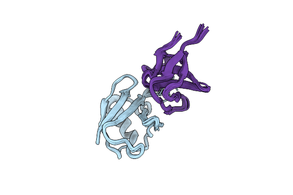
Distinct Ubiquitin Binding Modes Exhibited By Sh3 Domains: Molecular Determinants And Functional Implications
Organism: Mus musculus, Homo sapiens
Method: SOLUTION NMR Release Date: 2014-02-05 Classification: CELL CYCLE/SIGNALING PROTEIN |
Organism: Mus musculus, Homo sapiens
Method: SOLUTION NMR
Release Date: 2014-02-05
 |
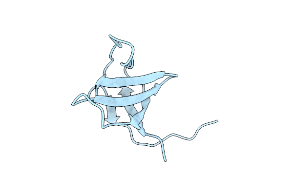
Distinct Ubiquitin Binding Modes Exhibited By Sh3 Domains: Molecular Determinants And Functional Implications
Organism: Homo sapiens, Mus musculus
Method: SOLUTION NMR Release Date: 2013-10-02 Classification: SIGNALING PROTEIN |
Organism: Homo sapiens, Mus musculus
Method: SOLUTION NMR
Release Date: 2013-10-02
 |
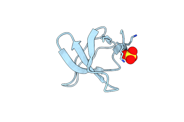
Crystal Structure Of Sh3C From Cin85
Organism: Homo sapiens
Method: X-RAY DIFFRACTION Resolution:2.05 Å Release Date: 2012-03-28 Classification: SIGNALING PROTEIN |
Organism: Homo sapiens
Method: X-RAY DIFFRACTION
Release Date: 2012-03-28
 |
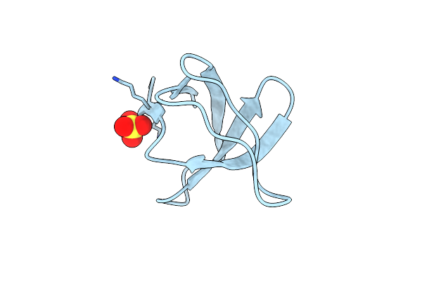
Crystal Structure Of The R21D Mutant Of Alpha-Spectrin Sh3 Domain. Crystal Obtained In Ammonium Sulphate At Ph 4.
Organism: Gallus gallus
Method: X-RAY DIFFRACTION Resolution:2.60 Å Release Date: 2011-01-19 Classification: SIGNALING PROTEIN Ligands: SO4 |
Organism: Gallus gallus
Method: X-RAY DIFFRACTION
Release Date: 2011-01-19
Ligands: SO4
 |
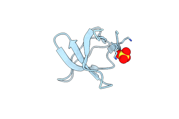
Crystal Structure Of The R21D Mutant Of Alpha-Spectrin Sh3 Domain. Crystal Obtained In Ammonium Sulphate At Ph 5.
Organism: Gallus gallus
Method: X-RAY DIFFRACTION Resolution:1.75 Å Release Date: 2011-01-19 Classification: SIGNALING PROTEIN Ligands: SO4 |
Organism: Gallus gallus
Method: X-RAY DIFFRACTION
Release Date: 2011-01-19
Ligands: SO4
 |
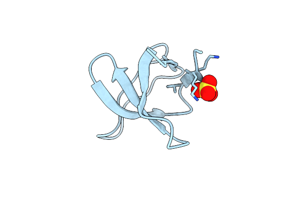
Crystal Structure Of The R21D Mutant Of Alpha-Spectrin Sh3 Domain. Crystal Obtained In Ammonium Sulphate At Ph 6.
Organism: Gallus gallus
Method: X-RAY DIFFRACTION Resolution:1.10 Å Release Date: 2011-01-19 Classification: SIGNALING PROTEIN Ligands: SO4 |
Organism: Gallus gallus
Method: X-RAY DIFFRACTION
Release Date: 2011-01-19
Ligands: SO4
 |
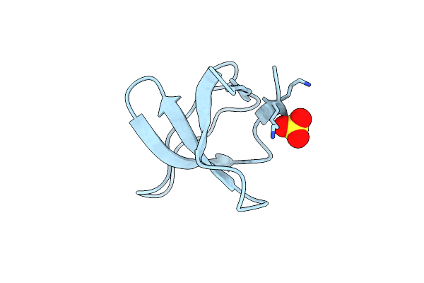
Crystal Structure Of The R21D Mutant Of Alpha-Spectrin Sh3 Domain. Crystal Obtained In Ammonium Sulphate At Ph 7
Organism: Gallus gallus
Method: X-RAY DIFFRACTION Resolution:1.60 Å Release Date: 2011-01-19 Classification: SIGNALING PROTEIN Ligands: SO4 |
Organism: Gallus gallus
Method: X-RAY DIFFRACTION
Release Date: 2011-01-19
Ligands: SO4
 |
Crystal Structure Of The R21D Mutant Of Alpha-Spectrin Sh3 Domain. Crystal Obtained In Ammonium Sulphate At Ph 9.
Organism: Gallus gallus
Method: X-RAY DIFFRACTION Resolution:1.70 Å Release Date: 2011-01-19 Classification: SIGNALING PROTEIN Ligands: SO4 |
Organism: Gallus gallus
Method: X-RAY DIFFRACTION
Release Date: 2011-01-19
Ligands: SO4