

Search Count: 20
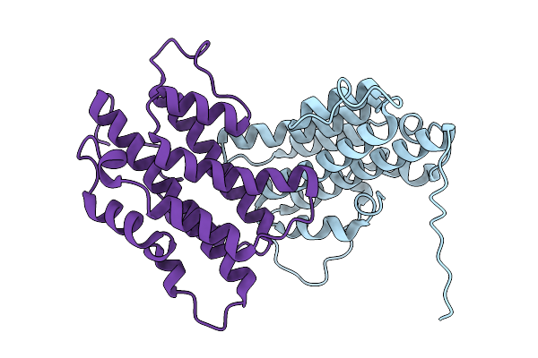 |
Structure Of S. Typhimurium 14028 Gifsy-1 Prophage Heps
Organism: Salmonella enterica subsp. enterica serovar typhimurium str. atcc 14028
Method: X-RAY DIFFRACTION Resolution:2.05 Å Release Date: 2026-01-28 Classification: ANTIVIRAL PROTEIN |
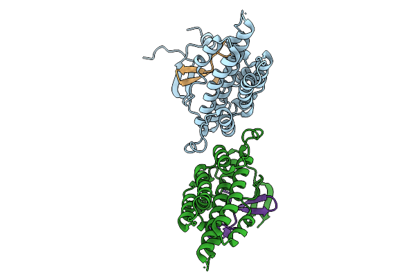 |
Structure Of S. Typhimurium 14028 Gifsy-1 Prophage Heps Bound To Bacteriophage Lambda J Tail Tip
Organism: Salmonella enterica subsp. enterica serovar typhimurium str. atcc 14028, Escherichia phage lambda
Method: X-RAY DIFFRACTION Resolution:1.86 Å Release Date: 2026-01-28 Classification: VIRAL PROTEIN |
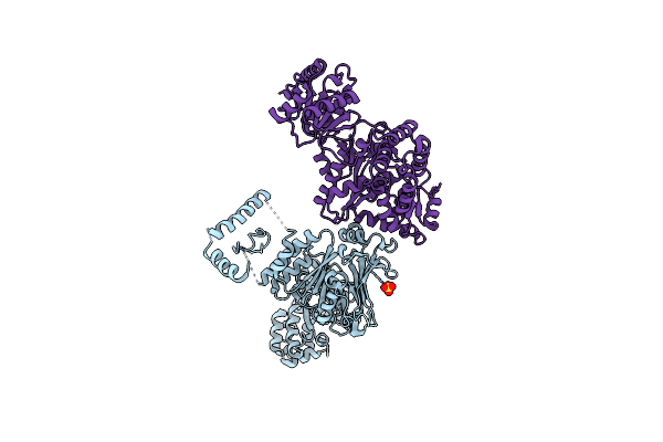 |
Structure Of Bacillus Cereus Vd045 Gabija Gaja-Gajb Complex
Organism: Bacillus cereus vd045
Method: X-RAY DIFFRACTION Resolution:3.00 Å Release Date: 2023-11-22 Classification: DNA BINDING PROTEIN/Hydrolase Ligands: SO4 |
Organism: Bacillus cereus vd045
Method: X-RAY DIFFRACTION
Release Date: 2023-11-22
Ligands: SO4
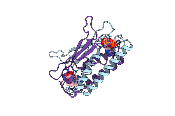 |
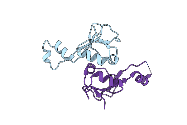
Structure Of Clostridium Botulinum Prophage Tad1 In Complex With 1''-3' Gcadpr
Organism: Clostridium botulinum
Method: X-RAY DIFFRACTION Resolution:2.10 Å Release Date: 2023-11-22 Classification: UNKNOWN FUNCTION Ligands: OJC |
Organism: Clostridium botulinum
Method: X-RAY DIFFRACTION
Release Date: 2023-11-22
Ligands: OJC
 |
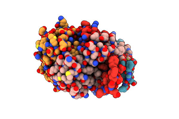
Structure Of Spo1 Phage Tad2 In Apo State
Organism: Okubovirus
Method: X-RAY DIFFRACTION Resolution:2.36 Å Release Date: 2023-11-22 Classification: VIRAL PROTEIN |
Organism: Okubovirus
Method: X-RAY DIFFRACTION
Release Date: 2023-11-22
 |
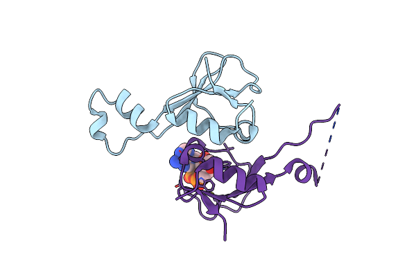
Structure Of Spo1 Phage Tad2 In Complex With 1''-3' Gcadpr
Organism: Okubovirus
Method: X-RAY DIFFRACTION Resolution:1.75 Å Release Date: 2023-11-22 Classification: VIRAL PROTEIN Ligands: OJC, MG |
Organism: Okubovirus
Method: X-RAY DIFFRACTION
Release Date: 2023-11-22
Ligands: OJC, MG
 |
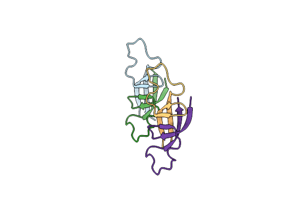
Structure Of Spo1 Phage Tad2 In Complex With 1''-2' Gcadpr
Organism: Okubovirus
Method: X-RAY DIFFRACTION Resolution:2.10 Å Release Date: 2023-11-22 Classification: VIRAL PROTEIN Ligands: MF6 |
Organism: Okubovirus
Method: X-RAY DIFFRACTION
Release Date: 2023-11-22
Ligands: MF6
 |
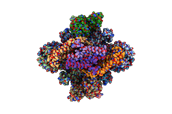
Structure Of Hachiman Anti-Defense 1 (Had1)
Organism: Unidentified
Method: X-RAY DIFFRACTION Resolution:2.00 Å Release Date: 2023-11-22 Classification: VIRAL PROTEIN |
Organism: Unidentified
Method: X-RAY DIFFRACTION
Release Date: 2023-11-22
 |
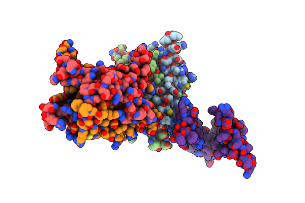
Structure Of The Phage Immune Evasion Protein Gad1 Bound To The Gabija Gajab Complex
Organism: Bacillus cereus vd045, Bacillus phage phi3t
Method: ELECTRON MICROSCOPY Release Date: 2023-11-22 Classification: VIRAL PROTEIN |
Organism: Bacillus cereus vd045, Bacillus phage phi3t
Method: ELECTRON MICROSCOPY
Release Date: 2023-11-22
 |
Structure Of Clostridium Botulinum Prophage Tad1 In Apo State
Organism: Clostridium botulinum
Method: X-RAY DIFFRACTION Resolution:2.20 Å Release Date: 2022-10-05 Classification: VIRAL PROTEIN |
Organism: Clostridium botulinum
Method: X-RAY DIFFRACTION
Release Date: 2022-10-05
 |
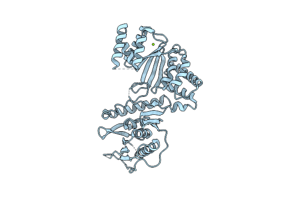
Structure Of Clostridium Botulinum Prophage Tad1 In Complex With 1''-2' Gcadpr
Organism: Clostridium botulinum
Method: X-RAY DIFFRACTION Resolution:1.72 Å Release Date: 2022-10-05 Classification: VIRAL PROTEIN Ligands: MF6 |
Organism: Clostridium botulinum
Method: X-RAY DIFFRACTION
Release Date: 2022-10-05
Ligands: MF6
 |
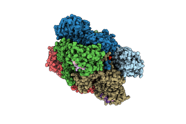
Structure Of Moraxella Osloensis Cap4 Saved/Carf-Domain Containing Receptor
Organism: Moraxella osloensis
Method: X-RAY DIFFRACTION Resolution:2.35 Å Release Date: 2020-06-17 Classification: HYDROLASE Ligands: MG |
Organism: Moraxella osloensis
Method: X-RAY DIFFRACTION
Release Date: 2020-06-17
Ligands: MG
 |
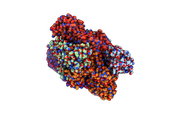
Structure Of Acinetobacter Baumannii Cap4 Saved/Carf-Domain Containing Receptor With The Cyclic Trinucleotide 2'3'3'-Caaa
Organism: Acinetobacter sp. atcc 27244, Synthetic construct
Method: X-RAY DIFFRACTION Resolution:2.10 Å Release Date: 2020-06-17 Classification: HYDROLASE Ligands: SO4 |
Organism: Acinetobacter sp. atcc 27244, Synthetic construct
Method: X-RAY DIFFRACTION
Release Date: 2020-06-17
Ligands: SO4
 |
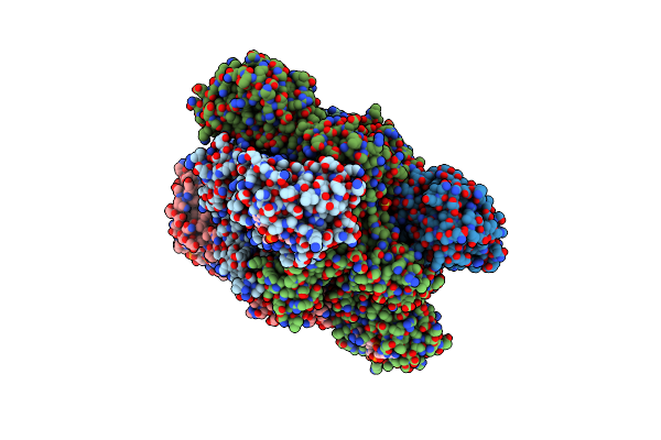
Structure Of Acinetobacter Baumannii Cap4 Saved/Carf-Domain Containing Receptor
Organism: Acinetobacter baumannii
Method: X-RAY DIFFRACTION Resolution:2.60 Å Release Date: 2020-06-17 Classification: HYDROLASE Ligands: SO4 |
Organism: Acinetobacter baumannii
Method: X-RAY DIFFRACTION
Release Date: 2020-06-17
Ligands: SO4
 |
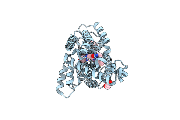
Structure Of Acinetobacter Baumannii Cap4 Saved/Carf-Domain Containing Receptor With The Cyclic Trinucleotide 3'3'3'-Caaa
Organism: Acinetobacter baumannii, Synthetic construct
Method: X-RAY DIFFRACTION Resolution:2.40 Å Release Date: 2020-06-17 Classification: HYDROLASE Ligands: SO4 |
Organism: Acinetobacter baumannii, Synthetic construct
Method: X-RAY DIFFRACTION
Release Date: 2020-06-17
Ligands: SO4
 |
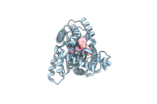
Catalytic Domain Of Human Phosphodiesterase 4B2B In Complex With A 5-Heterocycle Pyrazolopyridine Inhibitor
Organism: Homo sapiens
Method: X-RAY DIFFRACTION Resolution:2.42 Å Release Date: 2011-08-03 Classification: HYDROLASE/HYDROLASE INHIBITOR Ligands: ZN, MG, ARS, ZG1, GOL |
Organism: Homo sapiens
Method: X-RAY DIFFRACTION
Release Date: 2011-08-03
Ligands: ZN, MG, ARS, ZG1, GOL
 |
Catalytic Domain Of Human Phosphodiesterase 4B2B In Complex With A 5-Heterocycle Pyrazolopyridine Inhibitor
Organism: Homo sapiens
Method: X-RAY DIFFRACTION Resolution:2.00 Å Release Date: 2011-08-03 Classification: HYDROLASE/HYDROLASE INHIBITOR Ligands: ZN, MG, ARS, ZG2, GOL |
Organism: Homo sapiens
Method: X-RAY DIFFRACTION
Release Date: 2011-08-03
Ligands: ZN, MG, ARS, ZG2, GOL
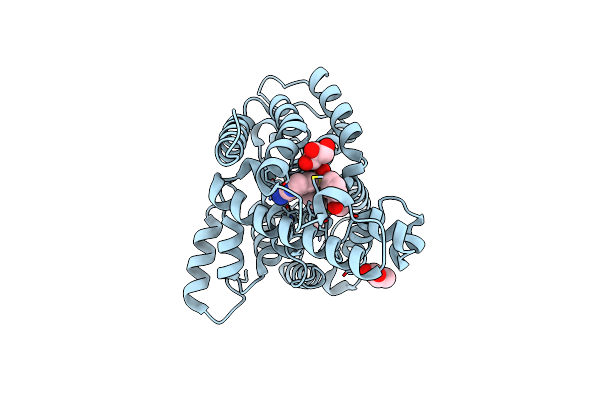 |
Catalytic Domain Of Human Phosphodiesterase 4B2B In Complex With A Quinoline Inhibitor
Organism: Homo sapiens
Method: X-RAY DIFFRACTION Resolution:1.75 Å Release Date: 2010-04-07 Classification: HYDROLASE Ligands: 066, ZN, MG, ARS, GOL |
Organism: Homo sapiens
Method: X-RAY DIFFRACTION
Release Date: 2010-04-07
Ligands: 066, ZN, MG, ARS, GOL
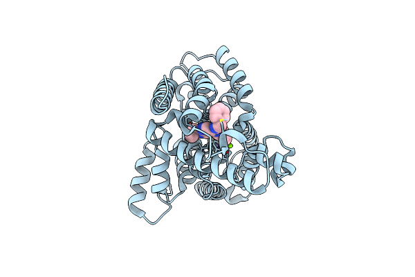 |
Crystal Structure Of Pde4B Catalytic Domain In Complex With A Pyrazolopyridine Inhibitor
Organism: Homo sapiens
Method: X-RAY DIFFRACTION Resolution:1.75 Å Release Date: 2009-05-19 Classification: HYDROLASE Ligands: ZN, MG, 20A, ARS |
Organism: Homo sapiens
Method: X-RAY DIFFRACTION
Release Date: 2009-05-19
Ligands: ZN, MG, 20A, ARS
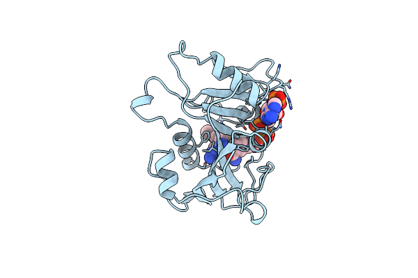 |
The Structure Of Pneumocystis Carinii Dihydrofolate Reductase To 1.9 Angstroms Resolution
Organism: Pneumocystis carinii
Method: X-RAY DIFFRACTION Resolution:1.86 Å Release Date: 1995-10-15 Classification: OXIDOREDUCTASE Ligands: NDP, TOP |
Organism: Pneumocystis carinii
Method: X-RAY DIFFRACTION
Release Date: 1995-10-15
Ligands: NDP, TOP