

Search Count: 3,123
 |
In Situ Structure Of The Side-By-Side Psii-Lhcii Dimer From Oryza Sativa (Composite)
Organism: Oryza sativa japonica group
Method: ELECTRON MICROSCOPY Resolution:3.50 Å Release Date: 2026-09-30 Classification: PHOTOSYNTHESIS Ligands: BCT |
Organism: Oryza sativa japonica group
Method: ELECTRON MICROSCOPY
Release Date: 2026-09-30
Ligands: BCT
 |
Grimophan Synthase From Variovorax Boronicumulans (Gris): Open Conformation
Organism: Variovorax boronicumulans
Method: X-RAY DIFFRACTION Resolution:2.40 Å Release Date: 2026-09-30 Classification: LYASE Ligands: PO4, PEG, EDO |
Organism: Variovorax boronicumulans
Method: X-RAY DIFFRACTION
Release Date: 2026-09-30
Ligands: PO4, PEG, EDO
 |
Chlororaphen Synthase From Pseudomonas Chlororaphis (Pc-Chlos) In Complex With Pyrophosphate
Organism: Pseudomonas chlororaphis
Method: X-RAY DIFFRACTION Resolution:2.00 Å Release Date: 2026-09-30 Classification: LYASE Ligands: MG, POP, GOL, BU3, EDO, ACT, NA |
Organism: Pseudomonas chlororaphis
Method: X-RAY DIFFRACTION
Release Date: 2026-09-30
Ligands: MG, POP, GOL, BU3, EDO, ACT, NA
 |
Chlororaphen Synthase From Pseudomonas Chlororaphis (Pc-Chlos) In Complex With Thiopyrophosphate And Bistris-Buffer
Organism: Pseudomonas chlororaphis
Method: X-RAY DIFFRACTION Resolution:1.25 Å Release Date: 2026-09-30 Classification: LYASE Ligands: MG, PIS, BTB, BU3, PEG, NA, EDO |
Organism: Pseudomonas chlororaphis
Method: X-RAY DIFFRACTION
Release Date: 2026-09-30
Ligands: MG, PIS, BTB, BU3, PEG, NA, EDO
 |
Chlororaphen Synthase From Pseudomonas Chlororaphis (Pc-Chlos) In Complex With Prechlororaphen Thiolopyrophosphate (A-Pcspp)
Organism: Pseudomonas chlororaphis
Method: X-RAY DIFFRACTION Resolution:1.90 Å Release Date: 2026-09-30 Classification: LYASE Ligands: MG, A1KN5, PEG, BU3, EDO, ACT, TRS, NA |
Organism: Pseudomonas chlororaphis
Method: X-RAY DIFFRACTION
Release Date: 2026-09-30
Ligands: MG, A1KN5, PEG, BU3, EDO, ACT, TRS, NA
 |
X-Ray Structure Of Human Psma Expressed In Sf9 Cells After Treatment With Endoglycosidase Endo-H
Organism: Homo sapiens
Method: X-RAY DIFFRACTION Resolution:2.60 Å Release Date: 2026-09-30 Classification: PEPTIDE BINDING PROTEIN Ligands: NAG, CL, CA, ZN, GOL |
Organism: Homo sapiens
Method: X-RAY DIFFRACTION
Release Date: 2026-09-30
Ligands: NAG, CL, CA, ZN, GOL
 |
Wild-Type Escherichia Coli Transhydrogenase Double Diiis Attached To Dii In The Presence Of Both Nadp+ And Nad+.
Organism: Escherichia coli k-12
Method: ELECTRON MICROSCOPY Release Date: 2026-09-30 Classification: MEMBRANE PROTEIN Ligands: PC1, NAP |
Organism: Escherichia coli k-12
Method: ELECTRON MICROSCOPY
Release Date: 2026-09-30
Ligands: PC1, NAP
 |
Wild-Type Escherichia Coli Transhydrogenase Single Diii Attached To Dii In The Presence Of Both Nadp+ And Nad+.
Organism: Escherichia coli k-12
Method: ELECTRON MICROSCOPY Release Date: 2026-09-30 Classification: MEMBRANE PROTEIN Ligands: NAP |
Organism: Escherichia coli k-12
Method: ELECTRON MICROSCOPY
Release Date: 2026-09-30
Ligands: NAP
 |
Wild-Type Escherichia Coli Transhydrogenase The Dissociated (Di)2 Dimer In The Presence Of Both Nadp+ And Nad+.
Organism: Escherichia coli k-12
Method: ELECTRON MICROSCOPY Release Date: 2026-09-30 Classification: MEMBRANE PROTEIN Ligands: NAD |
Organism: Escherichia coli k-12
Method: ELECTRON MICROSCOPY
Release Date: 2026-09-30
Ligands: NAD
 |
Escherichia Coli Nicotinamide Nucleotide Transhydrogenase Double Diiis Attached To Diis State In The Presence Of Nadph
Organism: Escherichia coli k-12
Method: ELECTRON MICROSCOPY Release Date: 2026-09-30 Classification: MEMBRANE PROTEIN Ligands: NDP |
Organism: Escherichia coli k-12
Method: ELECTRON MICROSCOPY
Release Date: 2026-09-30
Ligands: NDP
 |
Glucosylglycerol Phosphorylase Mutant-E231Q
Organism: Marinobacter salinexigens
Method: X-RAY DIFFRACTION Resolution:2.95 Å Release Date: 2026-09-30 Classification: HYDROLASE Ligands: GLC, PO4, MG, GOL |
Organism: Marinobacter salinexigens
Method: X-RAY DIFFRACTION
Release Date: 2026-09-30
Ligands: GLC, PO4, MG, GOL
 |
Wild Type Msggp Complex With Glucose
Organism: Marinobacter salinexigens
Method: X-RAY DIFFRACTION Resolution:2.90 Å Release Date: 2026-09-30 Classification: HYDROLASE Ligands: PEG, GOL, GLC, MG |
Organism: Marinobacter salinexigens
Method: X-RAY DIFFRACTION
Release Date: 2026-09-30
Ligands: PEG, GOL, GLC, MG
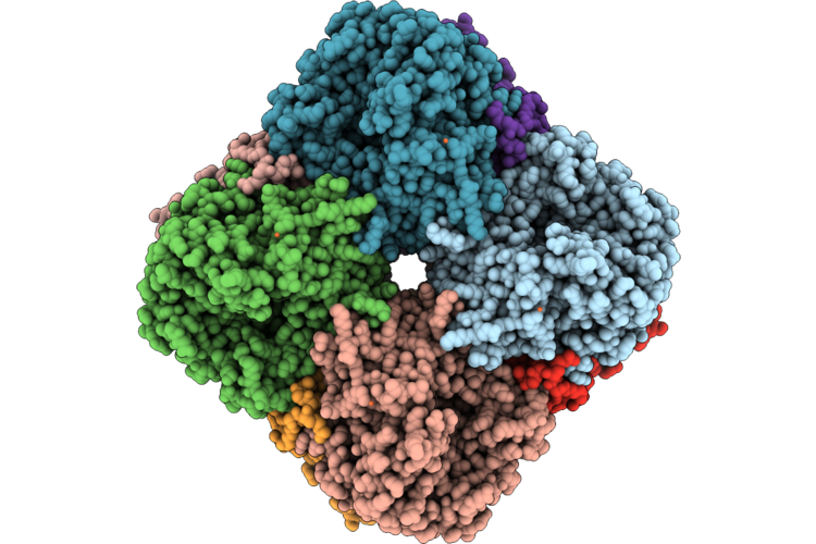 |
Cryoem Structure Of A Dntpase From Vibrio Cholerae
Organism: Vibrio cholerae o1 biovar el tor str. n16961
Method: ELECTRON MICROSCOPY Resolution:2.77 Å Release Date: 2026-09-23 Classification: ANTIMICROBIAL PROTEIN Ligands: MG, FE |
Organism: Vibrio cholerae o1 biovar el tor str. n16961
Method: ELECTRON MICROSCOPY
Release Date: 2026-09-23
Ligands: MG, FE
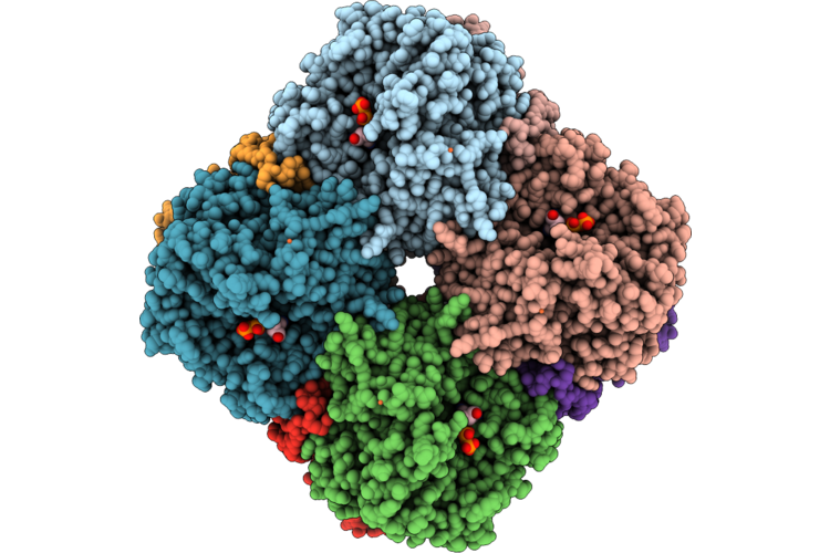 |
Cryoem Structure Of A Dntpase From Vibrio Cholerae With Dgtp
Organism: Vibrio cholerae serotype o1 (strain atcc 39315 / el tor inaba n16961)
Method: ELECTRON MICROSCOPY Resolution:2.70 Å Release Date: 2026-09-23 Classification: ANTIMICROBIAL PROTEIN Ligands: MG, DGT, FE |
Organism: Vibrio cholerae serotype o1 (strain atcc 39315 / el tor inaba n16961)
Method: ELECTRON MICROSCOPY
Release Date: 2026-09-23
Ligands: MG, DGT, FE
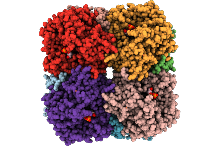 |
Cryoem Structure Of A Dntpase From Vibrio Cholerae With Gtp
Organism: Vibrio cholerae serotype o1 (strain atcc 39315 / el tor inaba n16961)
Method: ELECTRON MICROSCOPY Resolution:2.94 Å Release Date: 2026-09-23 Classification: ANTIMICROBIAL PROTEIN Ligands: MG, GTP, FE |
Organism: Vibrio cholerae serotype o1 (strain atcc 39315 / el tor inaba n16961)
Method: ELECTRON MICROSCOPY
Release Date: 2026-09-23
Ligands: MG, GTP, FE
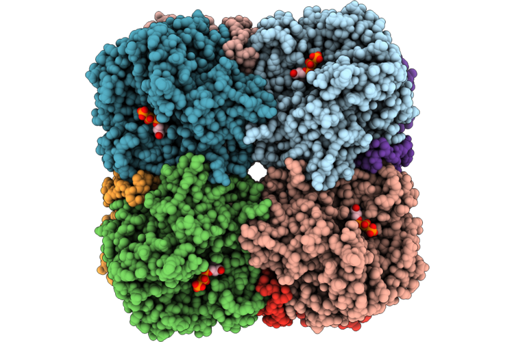 |
Cryoem Structure Of A Dntpase From Vibrio Cholerae With Gtp
Organism: Vibrio cholerae serotype o1 (strain atcc 39315 / el tor inaba n16961)
Method: ELECTRON MICROSCOPY Resolution:3.06 Å Release Date: 2026-09-23 Classification: ANTIMICROBIAL PROTEIN Ligands: MG, DGT |
Organism: Vibrio cholerae serotype o1 (strain atcc 39315 / el tor inaba n16961)
Method: ELECTRON MICROSCOPY
Release Date: 2026-09-23
Ligands: MG, DGT
 |
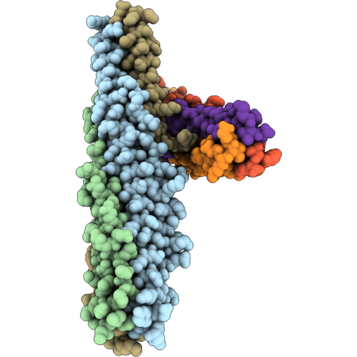
Pmcas12M-Crrna-Target Dna Complex
Organism: Pelomicrobium methylotrophicum
Method: ELECTRON MICROSCOPY Resolution:3.21 Å Release Date: 2026-09-23 Classification: DNA BINDING PROTEIN/RNA/DNA Ligands: MG |
Organism: Pelomicrobium methylotrophicum
Method: ELECTRON MICROSCOPY
Release Date: 2026-09-23
Ligands: MG
 |
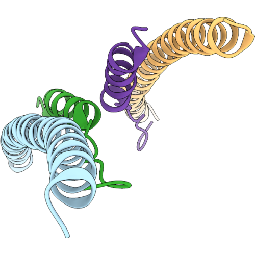
Crystal Structure Of The Sars-Cov-2 Spike Protein Hr1 In Complex With A Designed Peptide Inhibitor Dp-S2Fi-0089 At 3.1 Angstrom Resolution
Organism: Synthetic construct, Severe acute respiratory syndrome coronavirus 2
Method: X-RAY DIFFRACTION Resolution:3.10 Å Release Date: 2026-09-16 Classification: ANTIVIRAL PROTEIN |
Organism: Synthetic construct, Severe acute respiratory syndrome coronavirus 2
Method: X-RAY DIFFRACTION
Release Date: 2026-09-16
 |
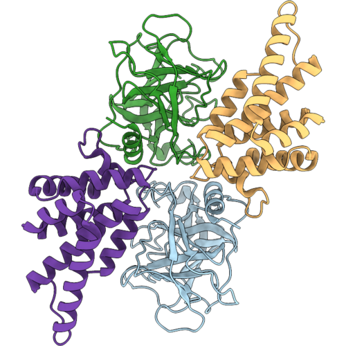
Crystal Structure Of The Sars-Cov-2 Spike Protein Hr1 In Complex With A Designed Peptide Inhibitor Dp-S2Fi-0087 At 3.1 Angstrom Resolution
Organism: Sars-cov-2 pseudovirus, Synthetic construct
Method: X-RAY DIFFRACTION Resolution:3.10 Å Release Date: 2026-09-16 Classification: ANTIVIRAL PROTEIN |
Organism: Sars-cov-2 pseudovirus, Synthetic construct
Method: X-RAY DIFFRACTION
Release Date: 2026-09-16
 |
Cryoem Structure Of Hgzma-Hgsdmbctd
Organism: Homo sapiens
Method: ELECTRON MICROSCOPY Resolution:3.59 Å Release Date: 2026-09-09 Classification: IMMUNE SYSTEM |
Organism: Homo sapiens
Method: ELECTRON MICROSCOPY
Release Date: 2026-09-09