

Search Count: 36
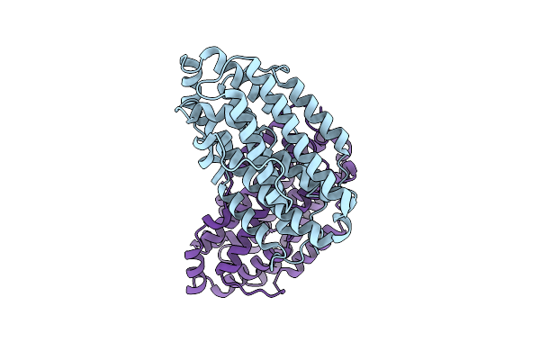 |
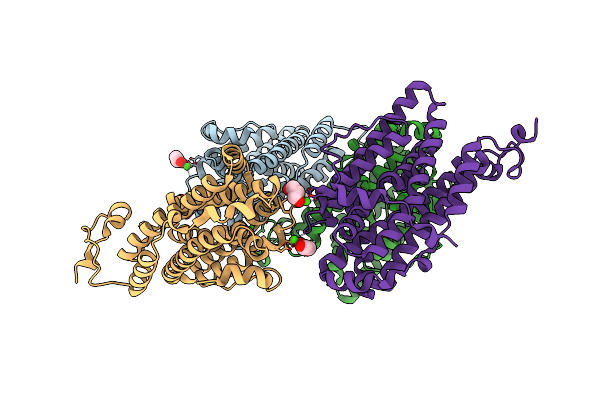
X-Ray Crystal Structure Of Francisella Hispaniensis Apo Ribonucleotide Reductase Beta Subunit
Organism: Francisella hispaniensis
Method: X-RAY DIFFRACTION Resolution:2.30 Å Release Date: 2025-09-17 Classification: OXIDOREDUCTASE Ligands: CL |
Organism: Francisella hispaniensis
Method: X-RAY DIFFRACTION
Release Date: 2025-09-17
Ligands: CL
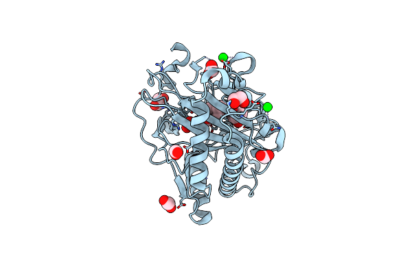 |
Structure Of Hyoscyamine 6-Beta Hydroxylase In Complex With Iron, 2-Oxoglutarate, And Hyoscyamine
Organism: Atropa belladonna
Method: X-RAY DIFFRACTION Resolution:1.53 Å Release Date: 2023-11-22 Classification: OXIDOREDUCTASE Ligands: SR, EDO, FMT, FE2, AKG, HYO |
Organism: Atropa belladonna
Method: X-RAY DIFFRACTION
Release Date: 2023-11-22
Ligands: SR, EDO, FMT, FE2, AKG, HYO
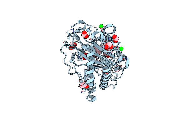 |
Structure Of Hyoscyamine 6-Beta Hydroxylase In Complex With Vanadyl, Succinate, And Hyoscyamine
Organism: Atropa belladonna
Method: X-RAY DIFFRACTION Resolution:1.79 Å Release Date: 2023-11-22 Classification: OXIDOREDUCTASE Ligands: EDO, HYO, SIN, FMT, V, O, SR |
Organism: Atropa belladonna
Method: X-RAY DIFFRACTION
Release Date: 2023-11-22
Ligands: EDO, HYO, SIN, FMT, V, O, SR
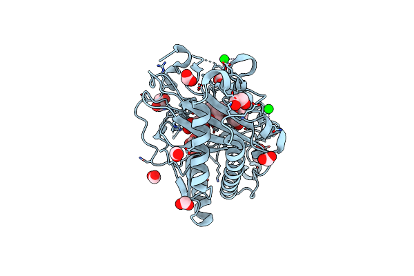 |
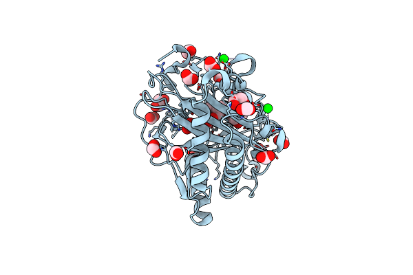
Structure Of Hyoscyamine 6-Beta Hydroxylase In Complex With Iron, Succinate, And 6-Oh-Hyoscyamine
Organism: Atropa belladonna
Method: X-RAY DIFFRACTION Resolution:1.58 Å Release Date: 2023-11-22 Classification: OXIDOREDUCTASE Ligands: FE2, EDO, FMT, OVR, SIN, SR |
Organism: Atropa belladonna
Method: X-RAY DIFFRACTION
Release Date: 2023-11-22
Ligands: FE2, EDO, FMT, OVR, SIN, SR
 |
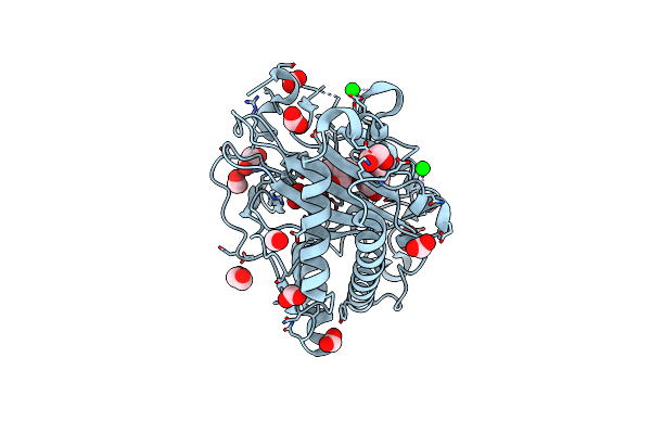
Structure Of Hyoscyamine 6-Beta Hydroxylase In Complex With Iron, 2-Oxoglutarate, And 6-Oh-Hyoscyamine
Organism: Atropa belladonna
Method: X-RAY DIFFRACTION Resolution:1.53 Å Release Date: 2023-11-22 Classification: OXIDOREDUCTASE Ligands: FE2, EDO, FMT, AKG, OVR, SR |
Organism: Atropa belladonna
Method: X-RAY DIFFRACTION
Release Date: 2023-11-22
Ligands: FE2, EDO, FMT, AKG, OVR, SR
 |
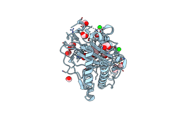
Structure Of Hyoscyamine 6-Beta Hydroxylase In Complex With Vanadyl, Succinate, And 6-Oh-Hyoscyamine
Organism: Atropa belladonna
Method: X-RAY DIFFRACTION Resolution:1.79 Å Release Date: 2023-11-22 Classification: OXIDOREDUCTASE Ligands: EDO, SIN, FMT, OVR, V, O, SR |
Organism: Atropa belladonna
Method: X-RAY DIFFRACTION
Release Date: 2023-11-22
Ligands: EDO, SIN, FMT, OVR, V, O, SR
 |
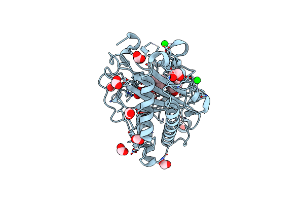
Structure Of Hyoscyamine 6-Beta Hydroxylase In Complex With Iron, Succinate, And Scopolamine
Organism: Atropa belladonna
Method: X-RAY DIFFRACTION Resolution:1.72 Å Release Date: 2023-11-22 Classification: OXIDOREDUCTASE Ligands: FE2, EDO, FMT, OW0, SIN, SR |
Organism: Atropa belladonna
Method: X-RAY DIFFRACTION
Release Date: 2023-11-22
Ligands: FE2, EDO, FMT, OW0, SIN, SR
 |
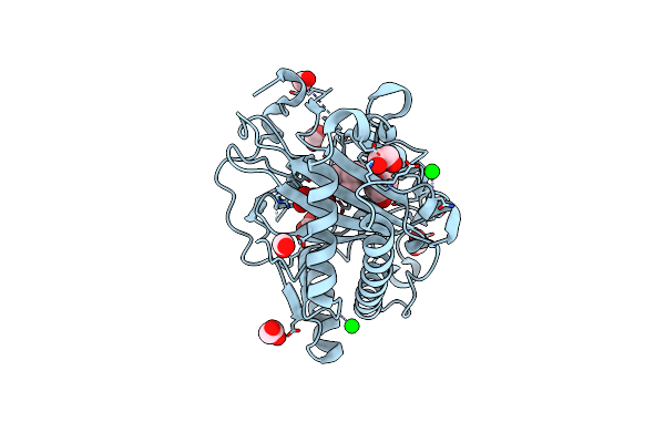
Structure Of L289F Hyoscyamine 6-Beta Hydroxylase In Complex With Iron, 2-Oxoglutarate, And Hyoscyamine
Organism: Atropa belladonna
Method: X-RAY DIFFRACTION Resolution:1.53 Å Release Date: 2023-11-22 Classification: OXIDOREDUCTASE Ligands: FE2, EDO, FMT, HYO, AKG, SR |
Organism: Atropa belladonna
Method: X-RAY DIFFRACTION
Release Date: 2023-11-22
Ligands: FE2, EDO, FMT, HYO, AKG, SR
 |
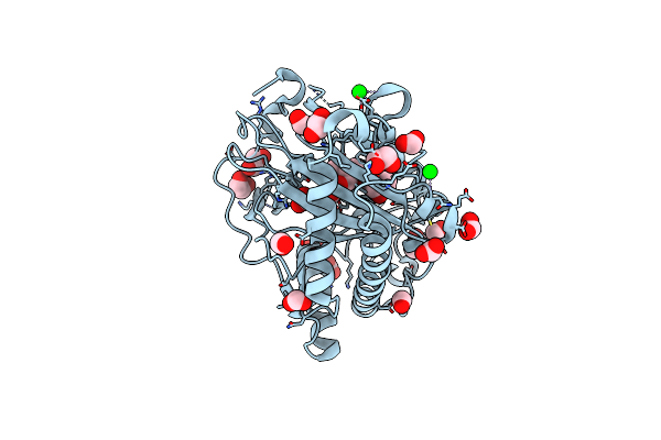
Structure Of L289F Hyoscyamine 6-Beta Hydroxylase In Complex With Vanadyl, Succinate, And Hyoscyamine
Organism: Atropa belladonna
Method: X-RAY DIFFRACTION Resolution:1.53 Å Release Date: 2023-11-22 Classification: OXIDOREDUCTASE Ligands: EDO, FMT, HYO, SIN, V, O, SR |
Organism: Atropa belladonna
Method: X-RAY DIFFRACTION
Release Date: 2023-11-22
Ligands: EDO, FMT, HYO, SIN, V, O, SR
 |
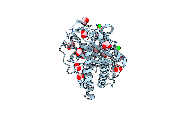
Structure Of L289F Hyoscyamine 6-Beta Hydroxylase In Complex With Iron, 2-Oxoglutarate, And 6-Oh-Hyoscyamine
Organism: Atropa belladonna
Method: X-RAY DIFFRACTION Resolution:1.56 Å Release Date: 2023-11-22 Classification: OXIDOREDUCTASE Ligands: FE2, EDO, FMT, AKG, OVR, SR |
Organism: Atropa belladonna
Method: X-RAY DIFFRACTION
Release Date: 2023-11-22
Ligands: FE2, EDO, FMT, AKG, OVR, SR
 |
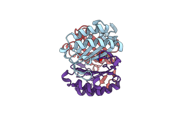
Structure Of L289F Hyoscyamine 6-Beta Hydroxylase In Complex With Vanadyl, Succinate, And 6-Oh-Hyoscyamine
Organism: Atropa belladonna
Method: X-RAY DIFFRACTION Resolution:2.03 Å Release Date: 2023-11-22 Classification: OXIDOREDUCTASE Ligands: EDO, FMT, SIN, OVR, V, SR |
Organism: Atropa belladonna
Method: X-RAY DIFFRACTION
Release Date: 2023-11-22
Ligands: EDO, FMT, SIN, OVR, V, SR
 |
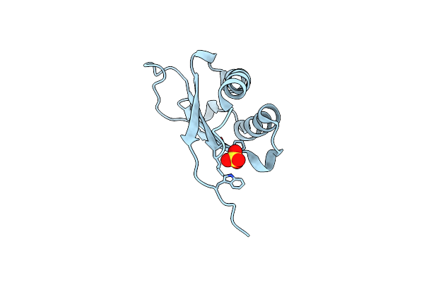
Crystal Structure Of S62A Mif2 Mutant
Organism: Homo sapiens
Method: X-RAY DIFFRACTION Resolution:1.33 Å Release Date: 2021-08-18 Classification: CYTOKINE Ligands: TRS |
Organism: Homo sapiens
Method: X-RAY DIFFRACTION
Release Date: 2021-08-18
Ligands: TRS
 |
F100A Mutant Structure Of Mif2 (D-Dt)
Organism: Homo sapiens
Method: X-RAY DIFFRACTION Resolution:1.57 Å Release Date: 2021-08-18 Classification: CYTOKINE Ligands: SO4 |
Organism: Homo sapiens
Method: X-RAY DIFFRACTION
Release Date: 2021-08-18
Ligands: SO4
 |
High-Resolution Crystal Structure Of Hmif2 With Tartrate At The Active Site
Organism: Homo sapiens
Method: X-RAY DIFFRACTION Resolution:1.27 Å Release Date: 2021-08-18 Classification: CYTOKINE Ligands: TLA |
Organism: Homo sapiens
Method: X-RAY DIFFRACTION
Release Date: 2021-08-18
Ligands: TLA
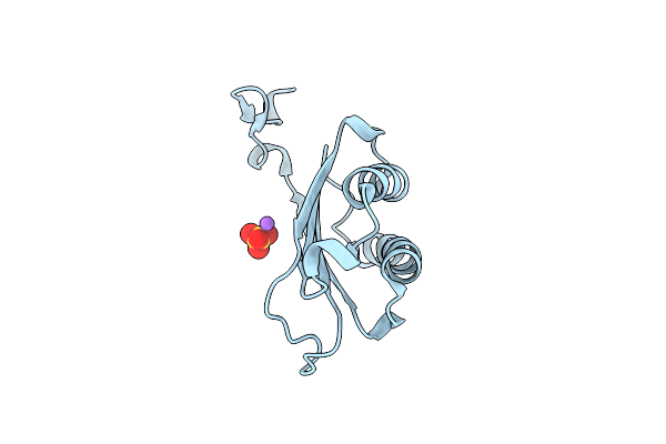 |
Crystal Structure Of P1G Mutant Of D-Dopachrome Tautomerase
Organism: Homo sapiens
Method: X-RAY DIFFRACTION Resolution:1.10 Å Release Date: 2021-08-18 Classification: CYTOKINE Ligands: SO4, NA |
Organism: Homo sapiens
Method: X-RAY DIFFRACTION
Release Date: 2021-08-18
Ligands: SO4, NA
 |
Pepducin Ut-Pep2 A Biased Allosteric Agonist Of Urotensin-Ii Receptor
Organism: Homo sapiens
Method: SOLUTION NMR Release Date: 2019-10-30 Classification: PEPTIDE BINDING PROTEIN |
Organism: Homo sapiens
Method: SOLUTION NMR
Release Date: 2019-10-30
 |
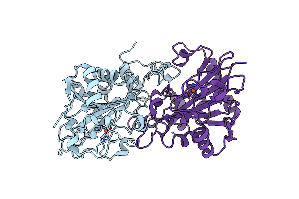
X-Ray Crystal Structure Of His-Tagged Y140F Ftmox1 Bound To Fe(Ii) And 2-Oxoglutarate
Organism: Neosartorya fumigata (strain atcc mya-4609 / af293 / cbs 101355 / fgsc a1100)
Method: X-RAY DIFFRACTION Resolution:1.92 Å Release Date: 2019-07-03 Classification: OXIDOREDUCTASE Ligands: FE2, AKG |
Organism: Neosartorya fumigata (strain atcc mya-4609 / af293 / cbs 101355 / fgsc a1100)
Method: X-RAY DIFFRACTION
Release Date: 2019-07-03
Ligands: FE2, AKG
 |
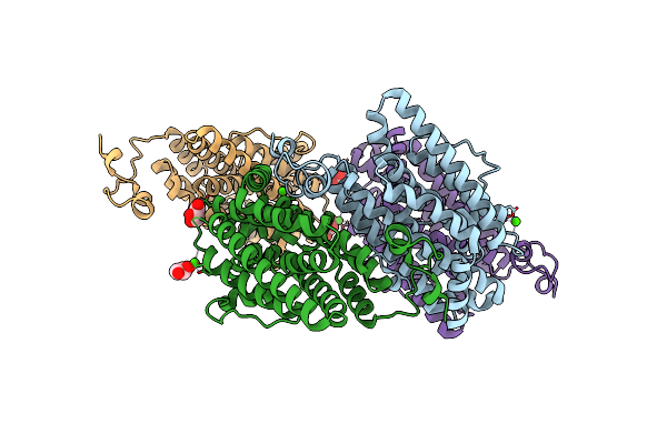
X-Ray Crystal Structure Of Y140F Ftmox1 Bound To Fe(Ii)
Organism: Neosartorya fumigata (strain atcc mya-4609 / af293 / cbs 101355 / fgsc a1100)
Method: X-RAY DIFFRACTION Resolution:1.55 Å Release Date: 2019-07-03 Classification: OXIDOREDUCTASE Ligands: FE2 |
Organism: Neosartorya fumigata (strain atcc mya-4609 / af293 / cbs 101355 / fgsc a1100)
Method: X-RAY DIFFRACTION
Release Date: 2019-07-03
Ligands: FE2
 |
Crystal Structure Of The Class Ie Ribonucleotide Reductase Beta Subunit From Aerococcus Urinae In Unactivated Form
Organism: Aerococcus urinae (strain acs-120-v-col10a)
Method: X-RAY DIFFRACTION Resolution:1.58 Å Release Date: 2018-09-19 Classification: OXIDOREDUCTASE Ligands: CA, GOL |
Organism: Aerococcus urinae (strain acs-120-v-col10a)
Method: X-RAY DIFFRACTION
Release Date: 2018-09-19
Ligands: CA, GOL
 |
Crystal Structure Of The Class Ie Ribonucleotide Reductase Beta Subunit From Aerococcus Urinae In Activated Form
Organism: Aerococcus urinae (strain acs-120-v-col10a)
Method: X-RAY DIFFRACTION Resolution:1.59 Å Release Date: 2018-09-19 Classification: OXIDOREDUCTASE Ligands: CA, GOL |
Organism: Aerococcus urinae (strain acs-120-v-col10a)
Method: X-RAY DIFFRACTION
Release Date: 2018-09-19
Ligands: CA, GOL