

Search Count: 126
 |
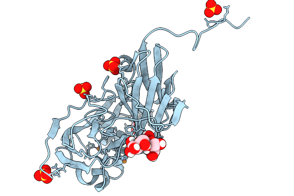
Atomic Resolution (1.02 A) Xfel Structure Of Nitrite-Bound Copper Nitrite Reductase From Bradyrhizobium Sp. Determined By Serial Femtosecond Rotation Crystallography (Sf-Rox) At 100 K
Organism: Bradyrhizobium diazoefficiens usda 110
Method: X-RAY DIFFRACTION Resolution:1.02 Å Release Date: 2026-03-25 Classification: OXIDOREDUCTASE Ligands: CU, NO2, GLC, FRU, SO4 |
Organism: Bradyrhizobium diazoefficiens usda 110
Method: X-RAY DIFFRACTION
Release Date: 2026-03-25
Ligands: CU, NO2, GLC, FRU, SO4
 |
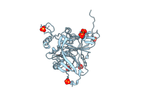
Atomic Resolution (1.00 A) Xfel Structure Of As-Isolated Copper Nitrite Reductase From Bradyrhizobium Sp. At High Ph (7.3) Determined By Serial Femtosecond Rotation Crystallography (Sf-Rox) At 100 K
Organism: Bradyrhizobium diazoefficiens usda 110
Method: X-RAY DIFFRACTION Resolution:1.00 Å Release Date: 2026-03-25 Classification: OXIDOREDUCTASE Ligands: CU, GLC, FRU, SO4 |
Organism: Bradyrhizobium diazoefficiens usda 110
Method: X-RAY DIFFRACTION
Release Date: 2026-03-25
Ligands: CU, GLC, FRU, SO4
 |
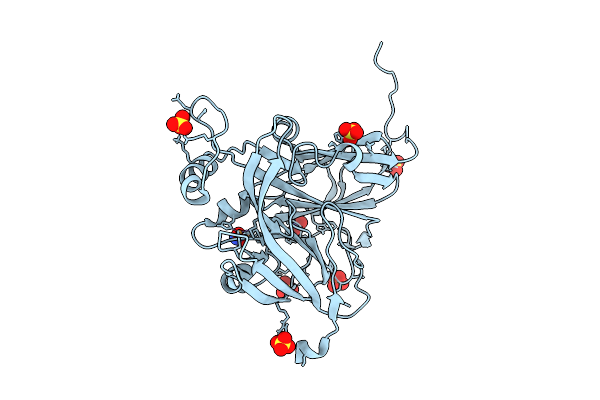
Sub-Atomic Resolution (0.95 A) Xfel Structure Of As-Isolated Copper Nitrite Reductase From Achromobacter Cycloclastes Determined By Serial Femtosecond Rotation Crystallography (Sf-Rox)
Organism: Achromobacter cycloclastes
Method: X-RAY DIFFRACTION Resolution:0.95 Å Release Date: 2026-03-18 Classification: OXIDOREDUCTASE Ligands: CU, SO4 |
Organism: Achromobacter cycloclastes
Method: X-RAY DIFFRACTION
Release Date: 2026-03-18
Ligands: CU, SO4
 |
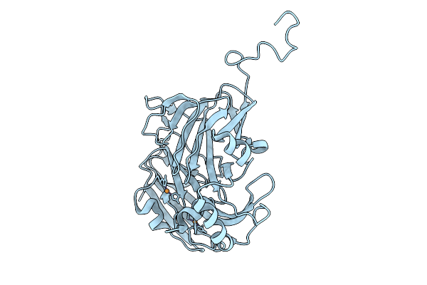
Sub-Atomic Resolution (0.95 A) Xfel Structure Of Nitrite-Bound Copper Nitrite Reductase From Achromobacter Cycloclastes Determined By Serial Femtosecond Rotation Crystallography (Sf-Rox) At 100 K
Organism: Achromobacter cycloclastes
Method: X-RAY DIFFRACTION Resolution:0.95 Å Release Date: 2026-03-18 Classification: OXIDOREDUCTASE Ligands: CU, SO4, NO2 |
Organism: Achromobacter cycloclastes
Method: X-RAY DIFFRACTION
Release Date: 2026-03-18
Ligands: CU, SO4, NO2
 |
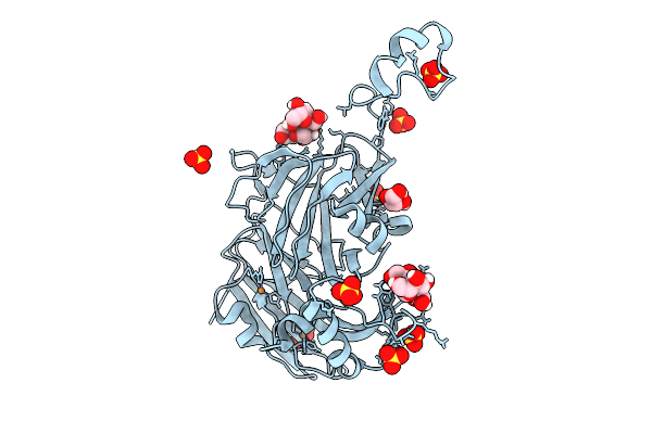
Atomic Resolution (1.05 A) Xfel Structure Of Chemically-Reduced Copper Nitrite Reductase From Bradyrhizobium Sp. Determined By Serial Femtosecond Rotation Crystallography (Sf-Rox) At 100 K
Organism: Bradyrhizobium sp. ors 375
Method: X-RAY DIFFRACTION Resolution:1.05 Å Release Date: 2026-03-18 Classification: OXIDOREDUCTASE Ligands: CU |
Organism: Bradyrhizobium sp. ors 375
Method: X-RAY DIFFRACTION
Release Date: 2026-03-18
Ligands: CU
 |
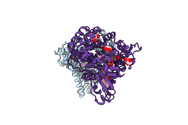
Atomic Resolution (1.15 A) Xfel Structure Of As-Isolated Copper Nitrite Reductase From Bradyrhizobium Sp. At Low Ph (5.5) Determined By Serial Femtosecond Rotation Crystallography (Sf-Rox) At 100 K
Organism: Bradyrhizobium sp. ors 375
Method: X-RAY DIFFRACTION Resolution:1.15 Å Release Date: 2026-03-18 Classification: OXIDOREDUCTASE Ligands: CU, GLC, FRU, SO4, GOL |
Organism: Bradyrhizobium sp. ors 375
Method: X-RAY DIFFRACTION
Release Date: 2026-03-18
Ligands: CU, GLC, FRU, SO4, GOL
 |
Synchrotron X-Ray Crystal Structure Of Oxygen-Bound F87A/F393H P450Bm3 With Decoy C7Prophe (N-Enanthyl-L-Prolyl-L-Phenylalanine) And Substrate Styrene At 2 Mgy X-Ray Dose
Organism: Priestia megaterium nbrc 15308 = atcc 14581
Method: X-RAY DIFFRACTION Resolution:1.60 Å Release Date: 2025-03-26 Classification: OXIDOREDUCTASE Ligands: HEM, SYN, OXY, D0L, GOL |
Organism: Priestia megaterium nbrc 15308 = atcc 14581
Method: X-RAY DIFFRACTION
Release Date: 2025-03-26
Ligands: HEM, SYN, OXY, D0L, GOL
 |
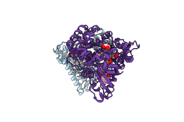
Xfel Crystal Structure Of The Oxidized Form Of F393H P450Bm3 With N-Enanthyl-L-Prolyl-L-Phenylalanine In Complex With Styrene
Organism: Priestia megaterium nbrc 15308 = atcc 14581
Method: X-RAY DIFFRACTION Resolution:1.80 Å Release Date: 2025-02-12 Classification: OXIDOREDUCTASE Ligands: HEM, D0L, SYN, GOL |
Organism: Priestia megaterium nbrc 15308 = atcc 14581
Method: X-RAY DIFFRACTION
Release Date: 2025-02-12
Ligands: HEM, D0L, SYN, GOL
 |
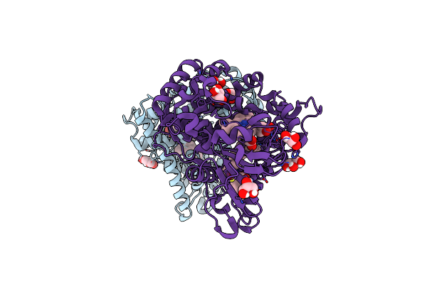
Xfel Crystal Structure Of The Oxidized Form Of F87A/F393H P450Bm3 With N-Enanthyl-L-Prolyl-L-Phenylalanine In Complex With Styrene
Organism: Priestia megaterium nbrc 15308 = atcc 14581
Method: X-RAY DIFFRACTION Resolution:1.85 Å Release Date: 2025-02-12 Classification: OXIDOREDUCTASE Ligands: HEM, D0L, SYN, GOL |
Organism: Priestia megaterium nbrc 15308 = atcc 14581
Method: X-RAY DIFFRACTION
Release Date: 2025-02-12
Ligands: HEM, D0L, SYN, GOL
 |
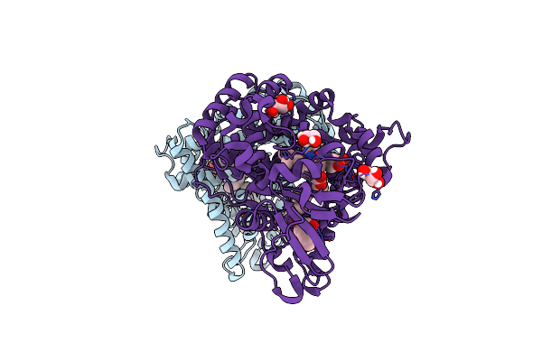
Xfel Crystal Structure Of The Reduced Form Of F393H P450Bm3 With N-Enanthyl-L-Prolyl-L-Phenylalanine In Complex With Styrene
Organism: Priestia megaterium nbrc 15308 = atcc 14581
Method: X-RAY DIFFRACTION Resolution:1.60 Å Release Date: 2025-02-12 Classification: OXIDOREDUCTASE Ligands: HEM, D0L, SYN, GOL |
Organism: Priestia megaterium nbrc 15308 = atcc 14581
Method: X-RAY DIFFRACTION
Release Date: 2025-02-12
Ligands: HEM, D0L, SYN, GOL
 |
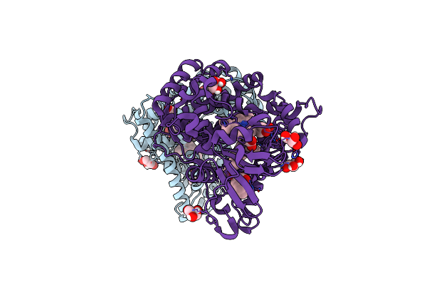
Xfel Crystal Structure Of The Reduced Form Of F87A/F393H P450Bm3 With N-Enanthyl-L-Prolyl-L-Phenylalanine In Complex With Styrene
Organism: Priestia megaterium nbrc 15308 = atcc 14581
Method: X-RAY DIFFRACTION Resolution:1.60 Å Release Date: 2025-02-12 Classification: OXIDOREDUCTASE Ligands: HEM, D0L, SYN, GOL |
Organism: Priestia megaterium nbrc 15308 = atcc 14581
Method: X-RAY DIFFRACTION
Release Date: 2025-02-12
Ligands: HEM, D0L, SYN, GOL
 |
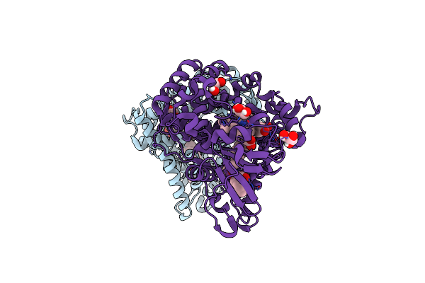
Xfel Crystal Structure Of The Oxygen-Bound Form Of F393H P450Bm3 With N-Enanthyl-L-Prolyl-L-Phenylalanine In Complex With Styrene
Organism: Priestia megaterium nbrc 15308 = atcc 14581
Method: X-RAY DIFFRACTION Resolution:1.50 Å Release Date: 2025-02-12 Classification: OXIDOREDUCTASE Ligands: HEM, D0L, SYN, OXY, GOL |
Organism: Priestia megaterium nbrc 15308 = atcc 14581
Method: X-RAY DIFFRACTION
Release Date: 2025-02-12
Ligands: HEM, D0L, SYN, OXY, GOL
 |
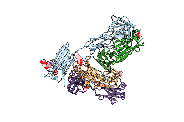
Xfel Crystal Structure Of The Oxygen-Bound Form Of F87A/F393H P450Bm3 With N-Enanthyl-L-Prolyl-L-Phenylalanine In Complex With Styrene
Organism: Priestia megaterium nbrc 15308 = atcc 14581
Method: X-RAY DIFFRACTION Resolution:1.50 Å Release Date: 2025-02-12 Classification: OXIDOREDUCTASE Ligands: HEM, D0L, SYN, OXY, GOL |
Organism: Priestia megaterium nbrc 15308 = atcc 14581
Method: X-RAY DIFFRACTION
Release Date: 2025-02-12
Ligands: HEM, D0L, SYN, OXY, GOL
 |
Crystal Structure Of Anti-Ige Antibody Hmk-12 Fab Complexed With Ige F(Ab')2
Organism: Rattus norvegicus, Mus musculus
Method: X-RAY DIFFRACTION Resolution:2.90 Å Release Date: 2024-07-31 Classification: IMMUNE SYSTEM Ligands: EDO, BOG |
Organism: Rattus norvegicus, Mus musculus
Method: X-RAY DIFFRACTION
Release Date: 2024-07-31
Ligands: EDO, BOG
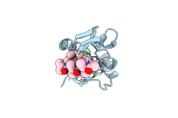 |
Mdm2 Bound With A Peptoid
Organism: Homo sapiens
Method: X-RAY DIFFRACTION Resolution:1.35 Å Release Date: 2024-05-01 Classification: LIGASE Ligands: UAC, CL |
Organism: Homo sapiens
Method: X-RAY DIFFRACTION
Release Date: 2024-05-01
Ligands: UAC, CL
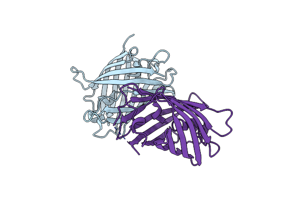 |
Crystal Structure Of A Highly Photostable And Bright Green Fluorescent Protein At Ph8.5
Organism: Synthetic construct
Method: X-RAY DIFFRACTION Resolution:1.56 Å Release Date: 2023-10-04 Classification: FLUORESCENT PROTEIN Ligands: CL |
Organism: Synthetic construct
Method: X-RAY DIFFRACTION
Release Date: 2023-10-04
Ligands: CL
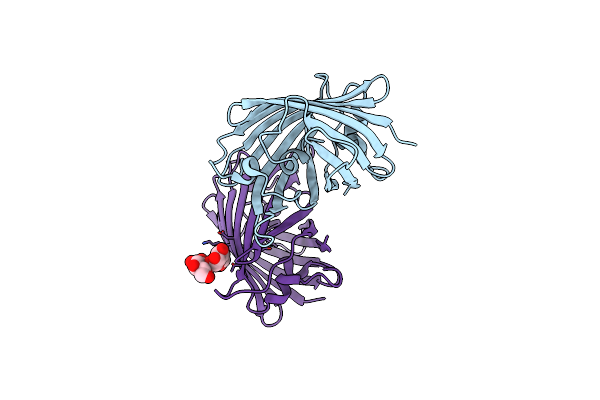 |
Crystal Structure Of A Highly Photostable And Bright Green Fluorescent Protein At Ph5.6
Organism: Synthetic construct
Method: X-RAY DIFFRACTION Resolution:2.20 Å Release Date: 2023-10-04 Classification: FLUORESCENT PROTEIN Ligands: CL |
Organism: Synthetic construct
Method: X-RAY DIFFRACTION
Release Date: 2023-10-04
Ligands: CL
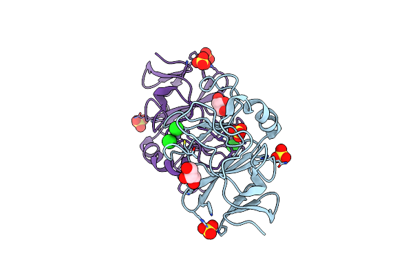 |
Crystal Structure Of The Immature Form Of Ttpeta
Organism: Thermochromatium tepidum
Method: X-RAY DIFFRACTION Resolution:1.70 Å Release Date: 2023-09-20 Classification: ELECTRON TRANSPORT Ligands: ZN, CL, SO4, GOL |
Organism: Thermochromatium tepidum
Method: X-RAY DIFFRACTION
Release Date: 2023-09-20
Ligands: ZN, CL, SO4, GOL
 |
Crystal Structure Of [2Fe-2S]-Ttpeta
Organism: Thermochromatium tepidum
Method: X-RAY DIFFRACTION Resolution:1.79 Å Release Date: 2023-09-20 Classification: ELECTRON TRANSPORT Ligands: FES, SO4, CL, GOL |
Organism: Thermochromatium tepidum
Method: X-RAY DIFFRACTION
Release Date: 2023-09-20
Ligands: FES, SO4, CL, GOL
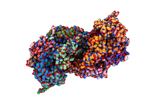 |
Re-Refinement Of Damage Free X-Ray Structure Of Bovine Cytochrome C Oxidase At 1.9 Angstrom Resolution
Organism: Bos taurus
Method: X-RAY DIFFRACTION Resolution:1.90 Å Release Date: 2022-07-20 Classification: OXIDOREDUCTASE Ligands: HEA, CU, MG, NA, PER, PGV, EDO, DMU, TGL, CUA, PSC, PEK, CDL, CHD, ZN, PO4 |