

Search Count: 10
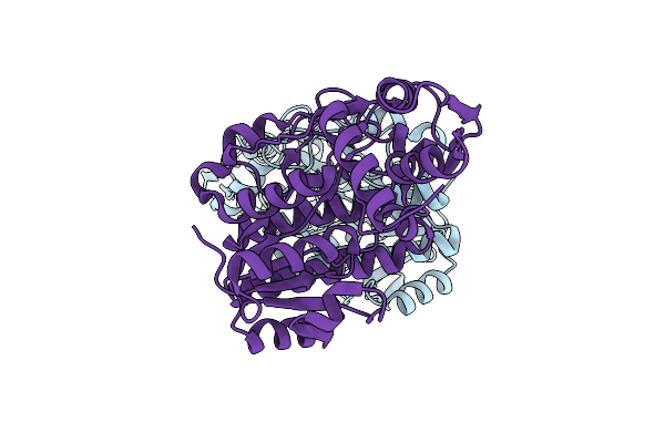 |
The X-Ray Structure Of L-Threonine Dehydrogenase From The Common Hospital Pathogen Clostridium Difficile.
Organism: Clostridioides difficile
Method: X-RAY DIFFRACTION Resolution:2.60 Å Release Date: 2021-06-16 Classification: OXIDOREDUCTASE |
Organism: Clostridioides difficile
Method: X-RAY DIFFRACTION
Release Date: 2021-06-16
 |
Three-Dimensional Structure Of L-Threonine 3-Dehydrogenase From Trypanosoma Brucei Refined To 1.77 Angstroms
Organism: Trypanosoma brucei
Method: X-RAY DIFFRACTION Resolution:1.77 Å Release Date: 2018-01-17 Classification: OXIDOREDUCTASE Ligands: NAD, ACT, CL, NA, GOL |
Organism: Trypanosoma brucei
Method: X-RAY DIFFRACTION
Release Date: 2018-01-17
Ligands: NAD, ACT, CL, NA, GOL
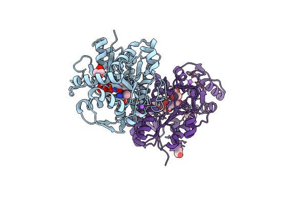 |
Three-Dimensional Structure Of L-Threonine 3-Dehydrogenase From Trypanosoma Brucei Bound To Nadh And L-Threonine Refined To 1.72 Angstroms
Organism: Trypanosoma brucei
Method: X-RAY DIFFRACTION Resolution:1.72 Å Release Date: 2018-01-10 Classification: OXIDOREDUCTASE Ligands: NAI, THR, GOL, NA |
Organism: Trypanosoma brucei
Method: X-RAY DIFFRACTION
Release Date: 2018-01-10
Ligands: NAI, THR, GOL, NA
 |
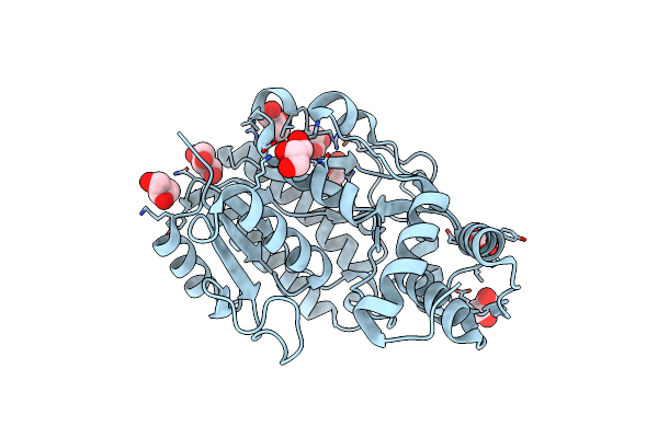
Three-Dimensional Structure Of L-Threonine 3-Dehydrogenase From Trypanosoma Brucei Bound To Nad+ Refined To 2.3 Angstroms
Organism: Trypanosoma brucei
Method: X-RAY DIFFRACTION Resolution:2.30 Å Release Date: 2017-11-15 Classification: OXIDOREDUCTASE Ligands: NAD, GOL, SO4 |
Organism: Trypanosoma brucei
Method: X-RAY DIFFRACTION
Release Date: 2017-11-15
Ligands: NAD, GOL, SO4
 |
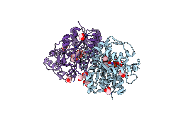
Three-Dimensional Structure Of L-Threonine 3-Dehydrogenase From Trypanosoma Brucei Refined To 2.1 Angstroms
Organism: Trypanosoma brucei
Method: X-RAY DIFFRACTION Resolution:2.10 Å Release Date: 2017-11-15 Classification: OXIDOREDUCTASE Ligands: GOL |
Organism: Trypanosoma brucei
Method: X-RAY DIFFRACTION
Release Date: 2017-11-15
Ligands: GOL
 |
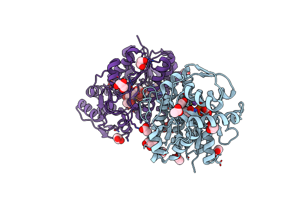
Three-Dimensional Structure Of L-Threonine 3-Dehydrogenase From Trypanosoma Brucei Showing Different Active Site Loop Conformations Between Dimer Subunits, Refined To 1.9 Angstroms
Organism: Trypanosoma brucei
Method: X-RAY DIFFRACTION Resolution:1.90 Å Release Date: 2017-11-15 Classification: OXIDOREDUCTASE Ligands: NAD, GOL, ACT |
Organism: Trypanosoma brucei
Method: X-RAY DIFFRACTION
Release Date: 2017-11-15
Ligands: NAD, GOL, ACT
 |
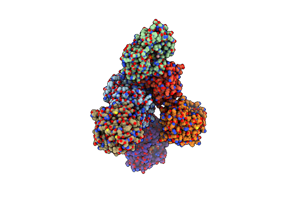
Three-Dimensional Structure Of L-Threonine 3-Dehydrogenase From Trypanosoma Brucei Bound To Nad+ Refined To 2.2 Angstroms
Organism: Trypanosoma brucei
Method: X-RAY DIFFRACTION Resolution:2.20 Å Release Date: 2017-11-15 Classification: OXIDOREDUCTASE Ligands: NAD, ACT, GOL, NA |
Organism: Trypanosoma brucei
Method: X-RAY DIFFRACTION
Release Date: 2017-11-15
Ligands: NAD, ACT, GOL, NA
 |
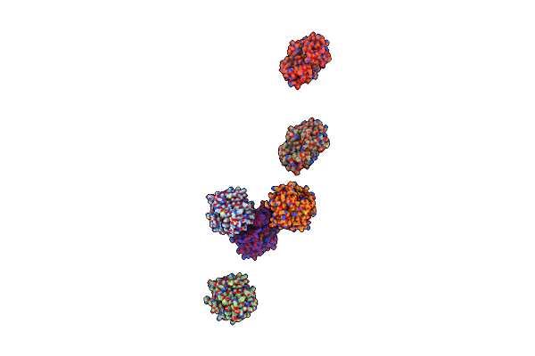
Three-Dimensional Structure Of L-Threonine 3-Dehydrogenase From Trypanosoma Brucei Bound To Nad+ And L-Allo-Threonine Refined To 2.23 Angstroms
Organism: Trypanosoma brucei
Method: X-RAY DIFFRACTION Resolution:2.26 Å Release Date: 2017-11-15 Classification: OXIDOREDUCTASE Ligands: NAD, GOL, ACT, ALO |
Organism: Trypanosoma brucei
Method: X-RAY DIFFRACTION
Release Date: 2017-11-15
Ligands: NAD, GOL, ACT, ALO
 |
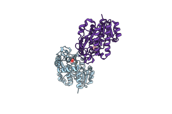
L-Threonine Dehydrogenase From Trypanosoma Brucei With Nad And The Inhibitor Pyruvate Bound.
Organism: Trypanosoma brucei
Method: X-RAY DIFFRACTION Resolution:2.10 Å Release Date: 2016-07-27 Classification: OXIDOREDUCTASE Ligands: NAD, PYR, BME, ACT, NA |
Organism: Trypanosoma brucei
Method: X-RAY DIFFRACTION
Release Date: 2016-07-27
Ligands: NAD, PYR, BME, ACT, NA
 |
L-Threonine Dehydrogenase From Trypanosoma Brucei.
Organism: Trypanosoma brucei
Method: X-RAY DIFFRACTION Resolution:1.45 Å Release Date: 2016-06-22 Classification: OXIDOREDUCTASE Ligands: ACT |
Organism: Trypanosoma brucei
Method: X-RAY DIFFRACTION
Release Date: 2016-06-22
Ligands: ACT