
Search Count: 84
All
Selected
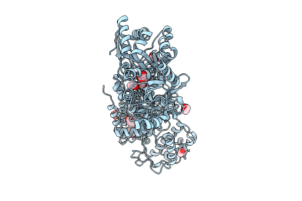 |
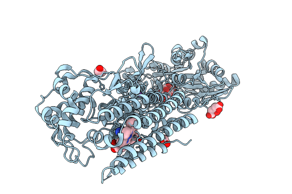
Crystal Structure Of The Indoleamine 2,3-Dioxygenagse 2 (Ido2) H143Y Mutant Complexed With 5-Methoxy-L-Trp
Organism: Homo sapiens
Method: X-RAY DIFFRACTION Resolution:2.60 Å Release Date: 2026-04-08 Classification: OXIDOREDUCTASE Ligands: A1A2Y, EDO, HEM, CYN, NA |
 |
Crystal Structure Of The Indoleamine 2,3-Dioxygenagse 2 (Ido2) Complexed With 5-Hydroxy-L-Trp
Organism: Homo sapiens
Method: X-RAY DIFFRACTION Resolution:2.68 Å Release Date: 2026-04-08 Classification: OXIDOREDUCTASE Ligands: EDO, HEM, CYN, 4PQ, NA |
 |
Crystal Structure Of The Indoleamine 2,3-Dioxygenagse 2 (Ido2) H143Y Mutant Complexed With 5-Methyl-L-Trp
Organism: Homo sapiens
Method: X-RAY DIFFRACTION Resolution:2.55 Å Release Date: 2026-04-08 Classification: OXIDOREDUCTASE Ligands: EDO, HEM, CYN, D0Q, NA |
 |
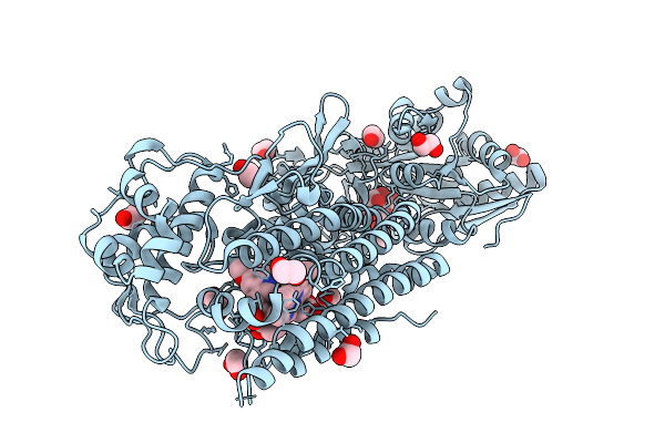
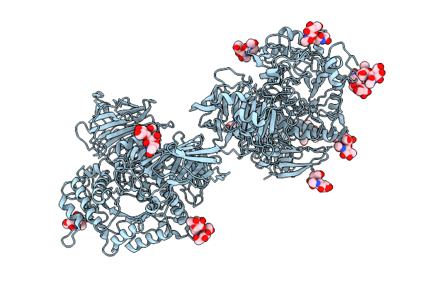
Crystal Structure Of The Indoleamine 2,3-Dioxygenagse 2 (Ido2) Complexed With L-Trp
Organism: Homo sapiens
Method: X-RAY DIFFRACTION Resolution:2.25 Å Release Date: 2026-04-08 Classification: OXIDOREDUCTASE Ligands: PEG, EDO, HEM, TRP, CYN, NA |
 |
Crystal Structure Of The Indoleamine 2,3-Dioxygenagse 2 (Ido2) Complexed With 5-Ht
Organism: Homo sapiens
Method: X-RAY DIFFRACTION Resolution:2.45 Å Release Date: 2026-04-08 Classification: OXIDOREDUCTASE Ligands: SRO, EDO, PEG, HEM, CYN, NA |
 |
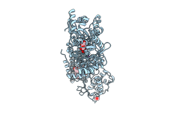
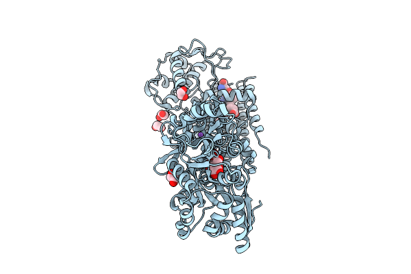
Crystal Structure Of The Indoleamine 2,3-Dioxygenagse 2 (Ido2) As Resting State
Organism: Homo sapiens
Method: X-RAY DIFFRACTION Resolution:2.45 Å Release Date: 2026-04-08 Classification: OXIDOREDUCTASE Ligands: EDO, CIT, HEM, NA |
 |
Crystal Structure Of The Indoleamine 2,3-Dioxygenagse 2 (Ido2) H143Y Mutant Complexed With L-Trp
Organism: Homo sapiens
Method: X-RAY DIFFRACTION Resolution:2.35 Å Release Date: 2026-04-08 Classification: OXIDOREDUCTASE Ligands: TRP, PEG, CIT, EDO, HEM, CYN, NA |
 |
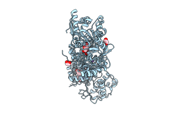
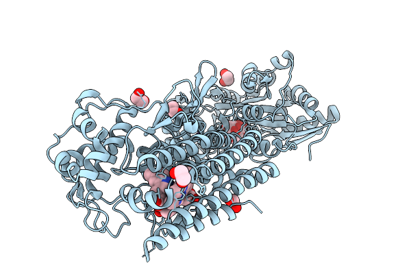
Crystal Structure Of The Indoleamine 2,3-Dioxygenagse 2 (Ido2) Complexed With Cyanide Ion
Organism: Homo sapiens
Method: X-RAY DIFFRACTION Resolution:2.55 Å Release Date: 2026-04-08 Classification: OXIDOREDUCTASE Ligands: CIT, EDO, HEM, CYN, NA |
 |
Crystal Structure Of The Indoleamine 2,3-Dioxygenagse 2 (Ido2) Complexed With D-Trp
Organism: Homo sapiens
Method: X-RAY DIFFRACTION Resolution:2.50 Å Release Date: 2026-04-08 Classification: OXIDOREDUCTASE Ligands: HEM, CYN, DTR, EDO, NA |
 |
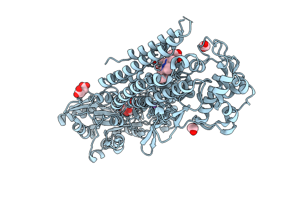
Crystal Structure Of The Indoleamine 2,3-Dioxygenagse 2 (Ido2) Complexed With 5-Methoxy-L-Trp
Organism: Homo sapiens
Method: X-RAY DIFFRACTION Resolution:2.50 Å Release Date: 2026-04-08 Classification: OXIDOREDUCTASE Ligands: A1A2Y, EDO, HEM, CYN |
 |
Crystal Structure Of The Indoleamine 2,3-Dioxygenagse 2 (Ido2) Complexed With 5-Methyl-L-Trp
Organism: Homo sapiens
Method: X-RAY DIFFRACTION Resolution:2.50 Å Release Date: 2026-04-08 Classification: OXIDOREDUCTASE Ligands: EDO, HEM, CYN, D0Q, NA |
 |
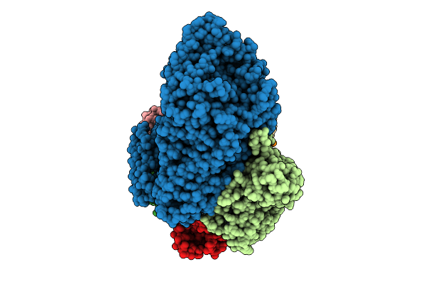
Organism: Sus scrofa domesticus
Method: ELECTRON MICROSCOPY Release Date: 2025-12-10 Classification: HYDROLASE Ligands: NAG |
 |
The Cryo-Em Structure Of Porcine Serum Mgam Bound With Acarviosyl-Maltotriose.
Organism: Sus scrofa domesticus
Method: ELECTRON MICROSCOPY Release Date: 2025-12-10 Classification: HYDROLASE Ligands: NAG |
 |
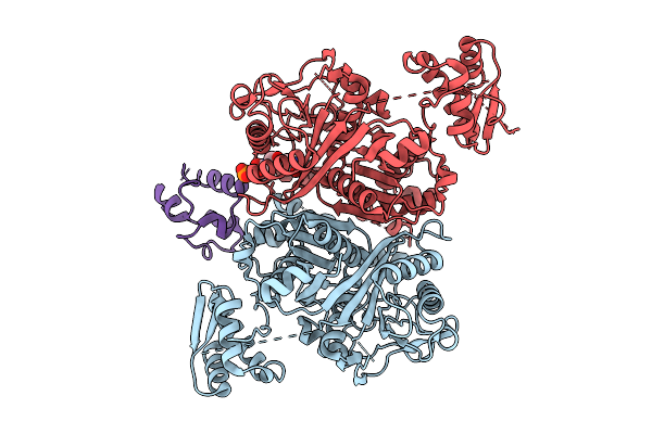
Organism: Archaeoglobus fulgidus dsm 4304, Pyrococcus furiosus dsm 3638, Synthetic construct
Method: ELECTRON MICROSCOPY Resolution:3.40 Å Release Date: 2025-12-10 Classification: RNA BINDING PROTEIN/RNA/DNA |
 |
Organism: Streptomyces graminofaciens, Synthetic construt
Method: X-RAY DIFFRACTION Resolution:2.00 Å Release Date: 2025-08-06 Classification: TRANSFERASE |
 |
Class 3 State Of The Gfsa Ksq-Ancestralat Chimeric Didomain In Complex With The Gfsa Acp Domain
Organism: Streptomyces graminofaciens
Method: ELECTRON MICROSCOPY Release Date: 2025-08-06 Classification: LYASE Ligands: 9EF |
 |
Class 1 State Of The Gfsa Ksq-Ancestralat Chimeric Didomain In Complex With The Gfsa Acp Domain
Organism: Streptomyces graminofaciens
Method: ELECTRON MICROSCOPY Release Date: 2025-08-06 Classification: LYASE Ligands: 9EF |
 |
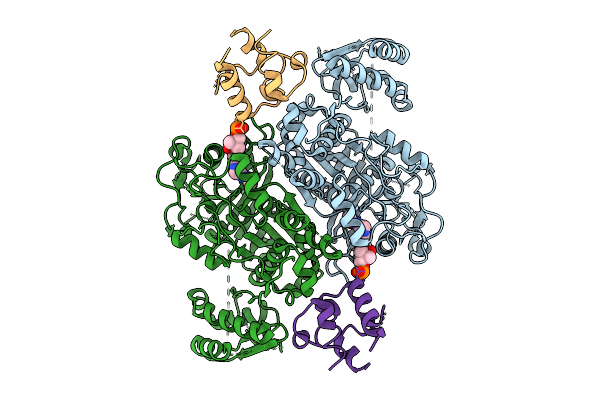
Organism: Bacillus sp. ps3
Method: ELECTRON MICROSCOPY Release Date: 2025-07-09 Classification: MEMBRANE PROTEIN Ligands: ADP, MG |
 |
Organism: Bacillus sp. ps3
Method: ELECTRON MICROSCOPY Release Date: 2025-07-09 Classification: MEMBRANE PROTEIN |
 |
Cryo-Em Structure Of The Inhibitor-Bound Vo Complex From Enterococcus Hirae
Organism: Enterococcus hirae atcc 9790
Method: ELECTRON MICROSCOPY Release Date: 2024-10-09 Classification: HYDROLASE/HYDROLASE INHIBITOR Ligands: CDL, NA, W3K |