

Search Count: 59
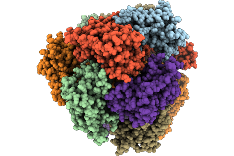 |
D0-State Of Wild Type Human Mitochondrial Lonp1 Protease Bound To Endogenous Adp
Organism: Homo sapiens
Method: ELECTRON MICROSCOPY Release Date: 2026-08-05 Classification: MOTOR PROTEIN Ligands: ADP |
Organism: Homo sapiens
Method: ELECTRON MICROSCOPY
Release Date: 2026-08-05
Ligands: ADP
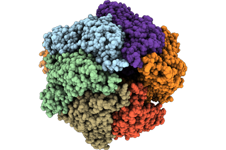 |
R0-State Of Wild Type Human Mitochondrial Lonp1 Protease Bound To Endogenous Adp
Organism: Homo sapiens
Method: ELECTRON MICROSCOPY Release Date: 2026-07-15 Classification: MOTOR PROTEIN Ligands: ADP |
Organism: Homo sapiens
Method: ELECTRON MICROSCOPY
Release Date: 2026-07-15
Ligands: ADP
 |
P2A-State Of Wild Type Human Mitochondrial Lonp1 Protease With Bound Substrate Protein, Adp And Aluminum Fluoride
Organism: Homo sapiens, Escherichia coli k-12
Method: ELECTRON MICROSCOPY Release Date: 2026-07-01 Classification: MOTOR PROTEIN Ligands: ADP, MG, AF3 |
Organism: Homo sapiens, Escherichia coli k-12
Method: ELECTRON MICROSCOPY
Release Date: 2026-07-01
Ligands: ADP, MG, AF3
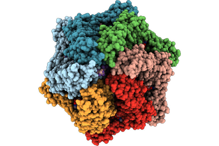 |
P1A-State Of Wild Type Human Mitochondrial Lonp1 Protease With Bound Substrate Protein, Adp And Aluminum Fluoride
Organism: Homo sapiens, Escherichia coli k-12
Method: ELECTRON MICROSCOPY Release Date: 2026-06-17 Classification: MOTOR PROTEIN Ligands: ADP, MG, AF3 |
Organism: Homo sapiens, Escherichia coli k-12
Method: ELECTRON MICROSCOPY
Release Date: 2026-06-17
Ligands: ADP, MG, AF3
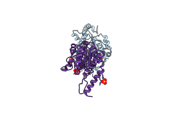 |
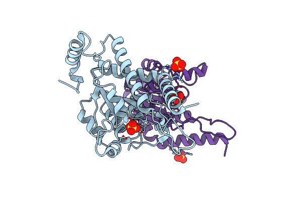
N-Acetylglucosamine 6-Phosphate Dehydratase: Inhibited 6-Phosphogluconic Acid State Of Nags
Organism: Streptomyces coelicolor
Method: X-RAY DIFFRACTION Resolution:1.69 Å Release Date: 2025-02-19 Classification: SUGAR BINDING PROTEIN Ligands: SO4, 6PG |
Organism: Streptomyces coelicolor
Method: X-RAY DIFFRACTION
Release Date: 2025-02-19
Ligands: SO4, 6PG
 |
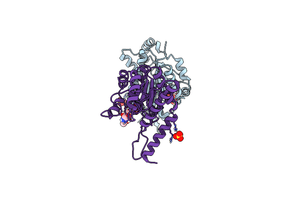
N-Acetylglucosamine 6-Phosphate Dehydratase: Apo Form Of Nags
Organism: Streptomyces coelicolor
Method: X-RAY DIFFRACTION Resolution:2.30 Å Release Date: 2025-02-19 Classification: SUGAR BINDING PROTEIN Ligands: SO4 |
Organism: Streptomyces coelicolor
Method: X-RAY DIFFRACTION
Release Date: 2025-02-19
Ligands: SO4
 |
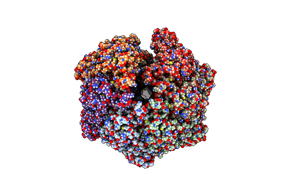
N-Acetylglucosamine 6-Phosphate Dehydratase: Glcnac6P Substrate-Bound State Of Nags
Organism: Streptomyces coelicolor
Method: X-RAY DIFFRACTION Resolution:2.59 Å Release Date: 2025-02-19 Classification: SUGAR BINDING PROTEIN Ligands: SO4, 16G |
Organism: Streptomyces coelicolor
Method: X-RAY DIFFRACTION
Release Date: 2025-02-19
Ligands: SO4, 16G
 |
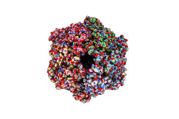
Human Lonp1, R-State, Incubated In Amppcp
Organism: Homo sapiens
Method: ELECTRON MICROSCOPY Release Date: 2021-12-22 Classification: MOTOR PROTEIN Ligands: ADP |
Organism: Homo sapiens
Method: ELECTRON MICROSCOPY
Release Date: 2021-12-22
Ligands: ADP
 |
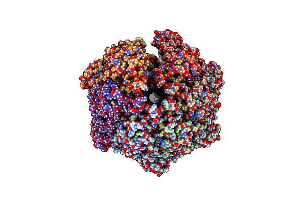
P2C-State Of Wild Type Human Mitochondrial Lonp1 Protease With Bound Endogenous Substrate Protein And In Presence Of Atp/Adp Mix
Organism: Homo sapiens
Method: ELECTRON MICROSCOPY Release Date: 2021-04-28 Classification: MOTOR PROTEIN Ligands: ATP, MG, ADP |
Organism: Homo sapiens
Method: ELECTRON MICROSCOPY
Release Date: 2021-04-28
Ligands: ATP, MG, ADP
 |
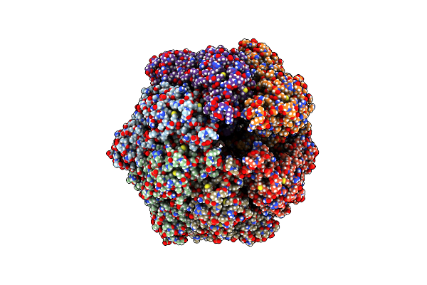
R-State Of Wild Type Human Mitochondrial Lonp1 Protease Bound To Endogenous Adp
Organism: Homo sapiens
Method: ELECTRON MICROSCOPY Release Date: 2021-04-28 Classification: MOTOR PROTEIN Ligands: ADP |
Organism: Homo sapiens
Method: ELECTRON MICROSCOPY
Release Date: 2021-04-28
Ligands: ADP
 |
D1-State Of Wild Type Human Mitochondrial Lonp1 Protease
Organism: Homo sapiens
Method: ELECTRON MICROSCOPY Release Date: 2021-04-28 Classification: MOTOR PROTEIN Ligands: ADP |
Organism: Homo sapiens
Method: ELECTRON MICROSCOPY
Release Date: 2021-04-28
Ligands: ADP
 |
Human Mitochondrial Lon Protease Homolog, D2-State
Organism: Homo sapiens
Method: ELECTRON MICROSCOPY Release Date: 2021-04-28 Classification: MOTOR PROTEIN Ligands: ADP |
Organism: Homo sapiens
Method: ELECTRON MICROSCOPY
Release Date: 2021-04-28
Ligands: ADP
 |
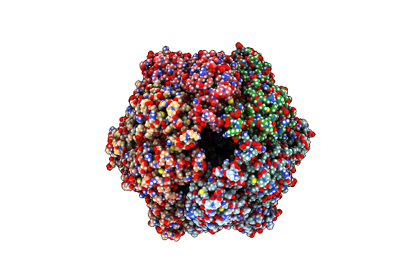
Amyloid Beta Oligomer Displayed On The Alpha Hemolysin Scaffold
Organism: Staphylococcus aureus, Homo sapiens
Method: ELECTRON MICROSCOPY Release Date: 2021-04-14 Classification: TOXIN |
Organism: Staphylococcus aureus, Homo sapiens
Method: ELECTRON MICROSCOPY
Release Date: 2021-04-14
 |
P2A-State Of Wild Type Human Mitochondrial Lonp1 Protease With Bound Substrate Protein And In Presence Of Atpgs
Organism: Homo sapiens
Method: ELECTRON MICROSCOPY Release Date: 2021-04-07 Classification: MOTOR PROTEIN Ligands: AGS, MG, ADP |
Organism: Homo sapiens
Method: ELECTRON MICROSCOPY
Release Date: 2021-04-07
Ligands: AGS, MG, ADP
 |
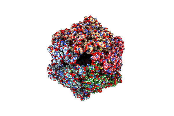
P1A-State Of Wild Type Human Mitochondrial Lonp1 Protease With Bound Substrate Protein And Atpgs
Organism: Homo sapiens
Method: ELECTRON MICROSCOPY Release Date: 2021-02-24 Classification: MOTOR PROTEIN Ligands: AGS, MG, ADP |
Organism: Homo sapiens
Method: ELECTRON MICROSCOPY
Release Date: 2021-02-24
Ligands: AGS, MG, ADP
 |
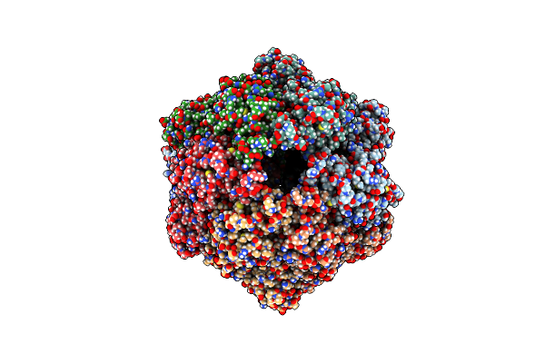
P1B-State Of Wild Type Human Mitochondrial Lonp1 Protease With Bound Endogenous Substrate Protein And In Presence Of Atp/Adp Mix
Organism: Homo sapiens
Method: ELECTRON MICROSCOPY Release Date: 2021-02-24 Classification: MOTOR PROTEIN Ligands: ATP, MG, ADP |
Organism: Homo sapiens
Method: ELECTRON MICROSCOPY
Release Date: 2021-02-24
Ligands: ATP, MG, ADP
 |
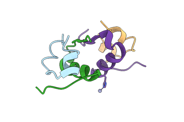
P1C-State Of Wild Type Human Mitochondrial Lonp1 Protease With Bound Substrate Protein In Presence Of Atp/Adp Mix
Organism: Homo sapiens
Method: ELECTRON MICROSCOPY Release Date: 2021-02-24 Classification: MOTOR PROTEIN Ligands: ATP, MG, ADP |
Organism: Homo sapiens
Method: ELECTRON MICROSCOPY
Release Date: 2021-02-24
Ligands: ATP, MG, ADP
 |
3D Electron Diffraction Structure Of Bovine Insulin
Organism: Bos taurus
Method: ELECTRON CRYSTALLOGRAPHY Resolution:3.25 Å Release Date: 2021-01-27 Classification: HORMONE Ligands: ZN |
Organism: Bos taurus
Method: ELECTRON CRYSTALLOGRAPHY
Release Date: 2021-01-27
Ligands: ZN
 |
3D Electron Diffraction Structure Of Thermolysin From Bacillus Thermoproteolyticus
Organism: Bacillus thermoproteolyticus
Method: ELECTRON CRYSTALLOGRAPHY Resolution:3.26 Å Release Date: 2021-01-27 Classification: HYDROLASE Ligands: ZN, CA |
Organism: Bacillus thermoproteolyticus
Method: ELECTRON CRYSTALLOGRAPHY
Release Date: 2021-01-27
Ligands: ZN, CA
 |
3D Electron Diffraction Structure Of Thaumatin From Thaumatococcus Daniellii
Organism: Thaumatococcus daniellii
Method: ELECTRON CRYSTALLOGRAPHY Resolution:2.76 Å Release Date: 2021-01-27 Classification: PLANT PROTEIN Ligands: CL |
Organism: Thaumatococcus daniellii
Method: ELECTRON CRYSTALLOGRAPHY
Release Date: 2021-01-27
Ligands: CL