

Search Count: 67
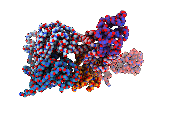 |
Intracellular Cryo-Tomography Structure Of Ebov Nucleocapsid At 8.9 Angstrom
Organism: Ebola virus - mayinga, zaire, 1976, Zaire
Method: ELECTRON MICROSCOPY Release Date: 2024-10-02 Classification: VIRAL PROTEIN |
Organism: Ebola virus - mayinga, zaire, 1976, Zaire
Method: ELECTRON MICROSCOPY
Release Date: 2024-10-02
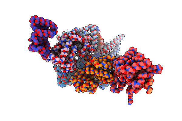 |
In-Virion Structure Of Ebola Virus Nucleocapsid-Like Assemblies From Recombinant Virus-Like Particles (Nucleoprotein, Vp24,Vp35,Vp40)
Organism: Ebola virus - mayinga, zaire, 1976, Zaire
Method: ELECTRON MICROSCOPY Release Date: 2024-10-02 Classification: VIRAL PROTEIN/RNA |
Organism: Ebola virus - mayinga, zaire, 1976, Zaire
Method: ELECTRON MICROSCOPY
Release Date: 2024-10-02
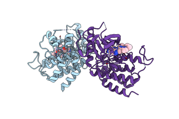 |
Fgfr2 Kinase Domain In Complex With A Pyrazolo[1,5-A]Pyrimidine Analog (Compound 29)
Organism: Homo sapiens
Method: X-RAY DIFFRACTION Resolution:2.68 Å Release Date: 2022-11-23 Classification: STRUCTURAL PROTEIN Ligands: U9P |
Organism: Homo sapiens
Method: X-RAY DIFFRACTION
Release Date: 2022-11-23
Ligands: U9P
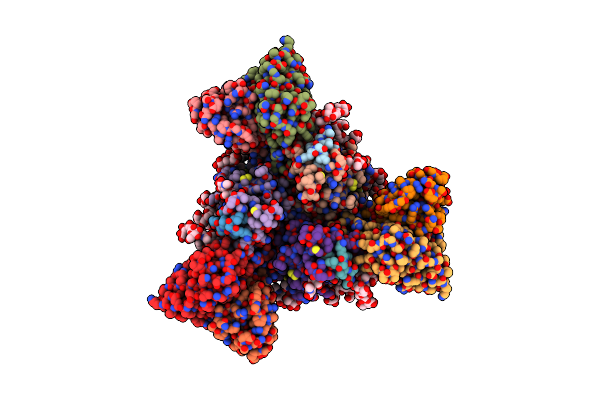 |
Native Lassa Glycoprotein In Complex With Neutralizing Antibodies 8.9F And 37.2D
Organism: Lassa virus, Homo sapiens
Method: ELECTRON MICROSCOPY Release Date: 2022-11-02 Classification: VIRAL PROTEIN/IMMUNE SYSTEM |
Organism: Lassa virus, Homo sapiens
Method: ELECTRON MICROSCOPY
Release Date: 2022-11-02
 |
Native Lassa Glycoprotein In Complex With Neutralizing Antibodies 12.1F And 37.2D
Organism: Homo sapiens, Lassa virus
Method: ELECTRON MICROSCOPY Release Date: 2022-11-02 Classification: VIRAL PROTEIN/IMMUNE SYSTEM Ligands: NAG |
Organism: Homo sapiens, Lassa virus
Method: ELECTRON MICROSCOPY
Release Date: 2022-11-02
Ligands: NAG
 |
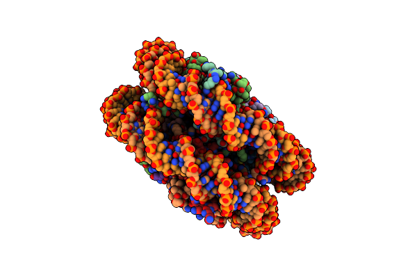
Insight Into The C-Terminal Sh3 Domain Mediated Binding Of Drosophila Drk To Sos And Dos
Organism: Drosophila melanogaster
Method: SOLUTION NMR Release Date: 2022-08-31 Classification: PEPTIDE BINDING PROTEIN |
Organism: Drosophila melanogaster
Method: SOLUTION NMR
Release Date: 2022-08-31
 |
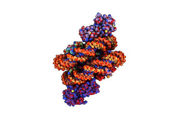
Chicken Knl2 In Complex With The Cenp-A Nucleosome
Organism: Gallus gallus, Homo sapiens, Synthetic construct
Method: ELECTRON MICROSCOPY Release Date: 2022-07-13 Classification: NUCLEAR PROTEIN/DNA |
Organism: Gallus gallus, Homo sapiens, Synthetic construct
Method: ELECTRON MICROSCOPY
Release Date: 2022-07-13
 |
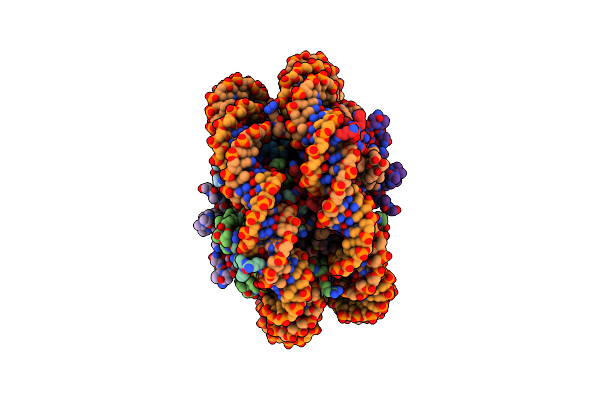
The Cryo-Em Structure Of Cenp-A Nucleosome In Complex With Cenp-C Peptide And Cenp-N N-Terminal Domain
Organism: Gallus gallus, Homo sapiens, Unidentified cloning vector, Synthetic construct
Method: ELECTRON MICROSCOPY Release Date: 2021-02-10 Classification: CELL CYCLE/DNA |
Organism: Gallus gallus, Homo sapiens, Unidentified cloning vector, Synthetic construct
Method: ELECTRON MICROSCOPY
Release Date: 2021-02-10
 |
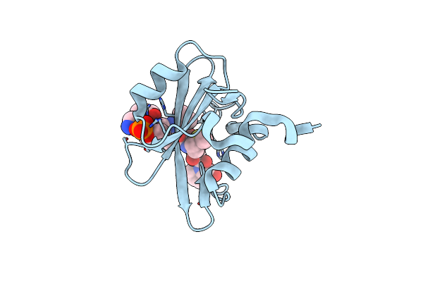
The Cryo-Em Structure Of Cenp-A Nucleosome In Complex With The Phosphorylated Cenp-C
Organism: Gallus gallus, Homo sapiens, Unidentified cloning vector, Synthetic construct
Method: ELECTRON MICROSCOPY Release Date: 2021-02-10 Classification: CELL CYCLE |
Organism: Gallus gallus, Homo sapiens, Unidentified cloning vector, Synthetic construct
Method: ELECTRON MICROSCOPY
Release Date: 2021-02-10
 |
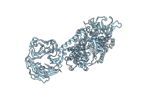
Molecular Interactions Of Cytoplasmic Region Of Ctla-4 With Sh2 Domains Of Pi3-Kinase
Organism: Homo sapiens
Method: X-RAY DIFFRACTION Resolution:1.10 Å Release Date: 2020-12-09 Classification: SIGNALING PROTEIN |
Organism: Homo sapiens
Method: X-RAY DIFFRACTION
Release Date: 2020-12-09
 |
Cryo-Em Structure Of The C-Terminal Half Of The Parkinson'S Disease-Linked Protein Leucine Rich Repeat Kinase 2 (Lrrk2)
Organism: Homo sapiens
Method: ELECTRON MICROSCOPY Release Date: 2020-08-26 Classification: SIGNALING PROTEIN Ligands: GDP, MG |
Organism: Homo sapiens
Method: ELECTRON MICROSCOPY
Release Date: 2020-08-26
Ligands: GDP, MG
 |
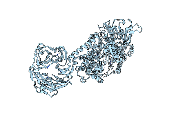
Cryo-Em Structure Of The C-Terminal Half Of The Parkinson'S Disease-Linked Protein Leucine Rich Repeat Kinase 2 (Lrrk2)
Organism: Homo sapiens
Method: ELECTRON MICROSCOPY Release Date: 2020-08-26 Classification: SIGNALING PROTEIN Ligands: GDP, MG |
Organism: Homo sapiens
Method: ELECTRON MICROSCOPY
Release Date: 2020-08-26
Ligands: GDP, MG
 |
Cryo-Em Structure Of The C-Terminal Half Of The Parkinson'S Disease-Linked Protein Leucine Rich Repeat Kinase 2 (Lrrk2)
Organism: Homo sapiens
Method: ELECTRON MICROSCOPY Release Date: 2020-08-26 Classification: SIGNALING PROTEIN |
Organism: Homo sapiens
Method: ELECTRON MICROSCOPY
Release Date: 2020-08-26
 |
Cryo-Em Structure Of The C-Terminal Half Of The Parkinson'S Disease-Linked Protein Leucine Rich Repeat Kinase 2 (Lrrk2)
Organism: Homo sapiens
Method: ELECTRON MICROSCOPY Release Date: 2020-08-26 Classification: SIGNALING PROTEIN |
Organism: Homo sapiens
Method: ELECTRON MICROSCOPY
Release Date: 2020-08-26
 |
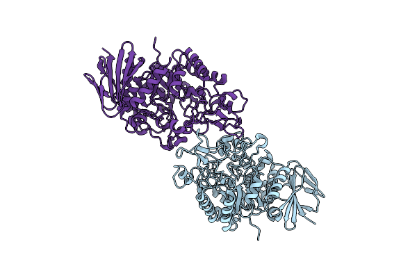
Integrative In Situ Structure Of Parkinsons Disease-Linked Human Lrrk2
Organism: Homo sapiens
Method: ELECTRON MICROSCOPY Release Date: 2020-08-19 Classification: SIGNALING PROTEIN, TRANSFERASE |
Organism: Homo sapiens
Method: ELECTRON MICROSCOPY
Release Date: 2020-08-19
 |
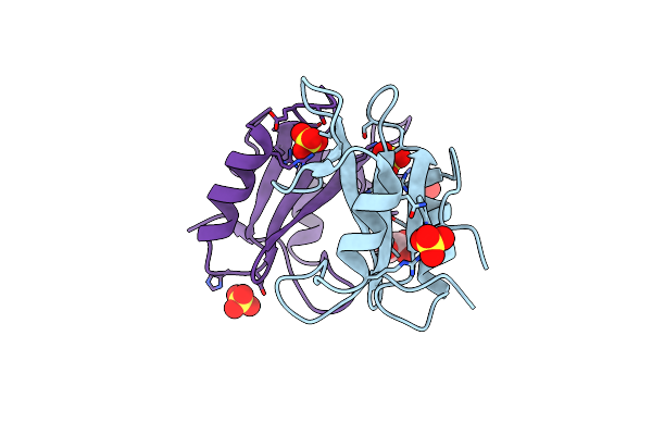
Crystal Structure Of Alpha-Glucosyl Transfer Enzyme, Xgta At 1.72 Angstrom Resolution
Organism: Xanthomonas campestris
Method: X-RAY DIFFRACTION Resolution:1.72 Å Release Date: 2019-07-24 Classification: HYDROLASE |
Organism: Xanthomonas campestris
Method: X-RAY DIFFRACTION
Release Date: 2019-07-24
 |
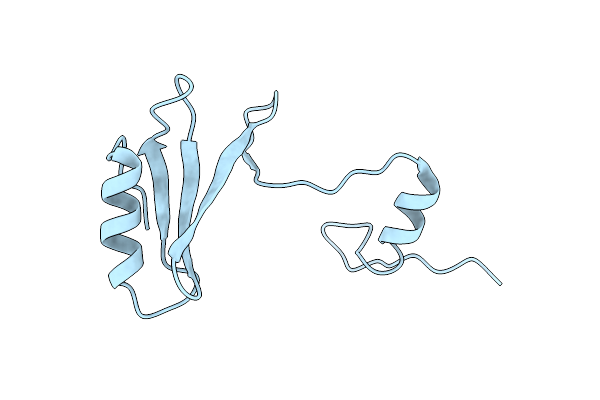
Grb2 Sh2 Domain In Phosphopeptide Free Form
Organism: Homo sapiens
Method: X-RAY DIFFRACTION Resolution:1.15 Å Release Date: 2019-07-17 Classification: SIGNALING PROTEIN Ligands: SO4, GOL |
Organism: Homo sapiens
Method: X-RAY DIFFRACTION
Release Date: 2019-07-17
Ligands: SO4, GOL
 |
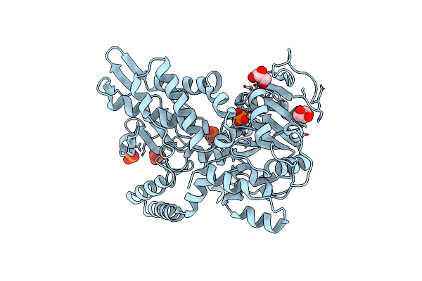
Grb2 Sh2 Domain In Domain Swapped Dimer Form
Organism: Homo sapiens
Method: X-RAY DIFFRACTION Resolution:2.00 Å Release Date: 2019-07-17 Classification: SIGNALING PROTEIN |
Organism: Homo sapiens
Method: X-RAY DIFFRACTION
Release Date: 2019-07-17
 |
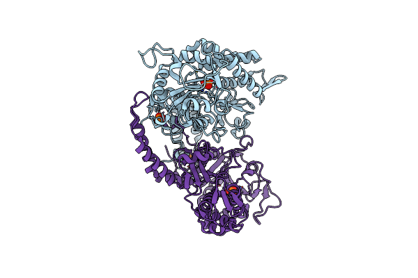
Crystal Structure Of The Double Mutant (D52N/D407A) Of Nt5C2-537X In The Active State
Organism: Homo sapiens
Method: X-RAY DIFFRACTION Resolution:1.98 Å Release Date: 2018-07-04 Classification: HYDROLASE Ligands: PO4, GOL |
Organism: Homo sapiens
Method: X-RAY DIFFRACTION
Release Date: 2018-07-04
Ligands: PO4, GOL
 |
Crystal Structure Of The Double Mutant (D52N/R367Q) Of Nt5C2-537X In The Basal State, Northeast Structural Genomics Consortium Target
Organism: Homo sapiens
Method: X-RAY DIFFRACTION Resolution:2.80 Å Release Date: 2018-07-04 Classification: HYDROLASE Ligands: MN, PO4 |
Organism: Homo sapiens
Method: X-RAY DIFFRACTION
Release Date: 2018-07-04
Ligands: MN, PO4