

Search Count: 242
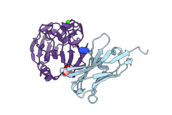 |
Crystal Structure Of The Complex Between Neuronal Pentraxin 2 (Np2 Ptx) And Antibody Fragment Vhh N1
Organism: Vicugna pacos, Homo sapiens
Method: X-RAY DIFFRACTION Resolution:2.02 Å Release Date: 2025-12-10 Classification: PROTEIN BINDING Ligands: AZI, CA, CL, GOL |
Organism: Vicugna pacos, Homo sapiens
Method: X-RAY DIFFRACTION
Release Date: 2025-12-10
Ligands: AZI, CA, CL, GOL
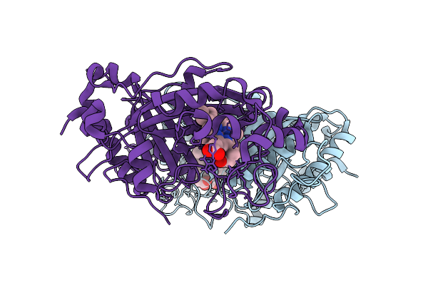 |
Dye Type Peroxidase Aa From Streptomyces Lividans With N3 Ligand By Serial Electron Diffraction (Serialed)
Organism: Streptomyces lividans
Method: ELECTRON CRYSTALLOGRAPHY Resolution:1.10 Å Release Date: 2025-07-16 Classification: OXIDOREDUCTASE Ligands: AZI, HEM |
Organism: Streptomyces lividans
Method: ELECTRON CRYSTALLOGRAPHY
Release Date: 2025-07-16
Ligands: AZI, HEM
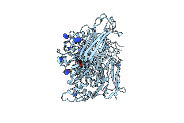 |
Cryo-Em Structure Of Thomasclavelia Ramosa Iga Peptidase (Igase) Active Site Mutant (S32-N876)
Organism: Thomasclavelia ramosa
Method: ELECTRON MICROSCOPY Release Date: 2025-06-25 Classification: HYDROLASE Ligands: ZN, GOL, AZI |
Organism: Thomasclavelia ramosa
Method: ELECTRON MICROSCOPY
Release Date: 2025-06-25
Ligands: ZN, GOL, AZI
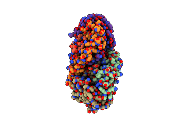 |
Crystal Structure Of The Soluble Green Pigment Protein From Tettigonia Cantans
Organism: Tettigonia cantans
Method: X-RAY DIFFRACTION Resolution:1.99 Å Release Date: 2025-04-23 Classification: TRANSPORT PROTEIN Ligands: LUT, AZI, PLC, A1L6M |
Organism: Tettigonia cantans
Method: X-RAY DIFFRACTION
Release Date: 2025-04-23
Ligands: LUT, AZI, PLC, A1L6M
 |
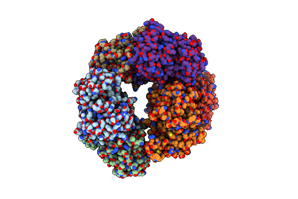
Crystal Structure Of Gh29 Family Alpha-L-Fucosidase From Fusarium Proliferatum Le1
Organism: Gibberella intermedia
Method: X-RAY DIFFRACTION Resolution:2.19 Å Release Date: 2025-03-19 Classification: HYDROLASE Ligands: NAG, TRS, MPD, NA, AZI, ACT |
Organism: Gibberella intermedia
Method: X-RAY DIFFRACTION
Release Date: 2025-03-19
Ligands: NAG, TRS, MPD, NA, AZI, ACT
 |
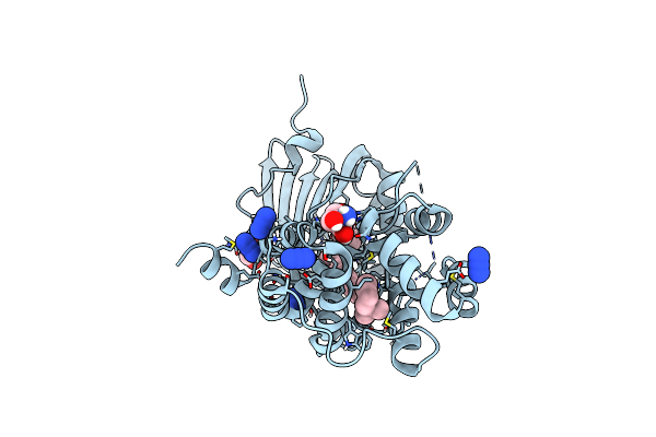
Binding Of Nadp To A Formate Dehydrogenase From Starkeya Novella.
Organism: Starkeya novella
Method: X-RAY DIFFRACTION Resolution:1.91 Å Release Date: 2024-04-24 Classification: STRUCTURAL PROTEIN Ligands: NAP, AZI |
Organism: Starkeya novella
Method: X-RAY DIFFRACTION
Release Date: 2024-04-24
Ligands: NAP, AZI
 |
Crystal Structure Of The Orange Carotenoid Protein 2 (Ocp2) From Gloeocapsa Sp. Pcc 7428
Organism: Gloeocapsa sp. pcc 7428
Method: X-RAY DIFFRACTION Resolution:1.80 Å Release Date: 2024-01-03 Classification: PHOTOSYNTHESIS Ligands: ECH, TRS, AZI, GOL |
Organism: Gloeocapsa sp. pcc 7428
Method: X-RAY DIFFRACTION
Release Date: 2024-01-03
Ligands: ECH, TRS, AZI, GOL
 |
Crystal Structure Of The Chimera Of Human 14-3-3 Zeta And Phosphorylated Cytoplasmic Loop Fragment Of The Alpha7 Acetylcholine Receptor
Organism: Homo sapiens
Method: X-RAY DIFFRACTION Resolution:1.95 Å Release Date: 2023-10-18 Classification: SIGNALING PROTEIN Ligands: EDO, AZI, BEZ, PEG |
Organism: Homo sapiens
Method: X-RAY DIFFRACTION
Release Date: 2023-10-18
Ligands: EDO, AZI, BEZ, PEG
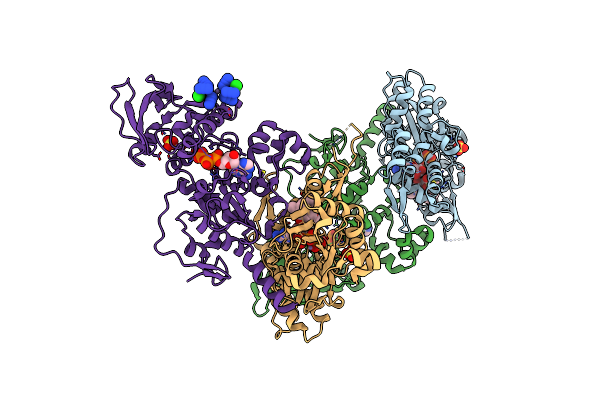 |
Exoy Nucleotidyl Cyclase Domain From Vibrio Nigripulchritudo Martx Toxin, Bound To Latrunculin-B-Atp-Mg-Actin, And 3'-Deoxyadenosine-5'-Triphosphate And 2 Mg Ions
Organism: Vibrio nigripulchritudo, Oryctolagus cuniculus
Method: X-RAY DIFFRACTION Resolution:2.50 Å Release Date: 2023-09-20 Classification: TOXIN Ligands: LAB, ATP, MG, SO4, 3AT, MN, AZI, CL |
Organism: Vibrio nigripulchritudo, Oryctolagus cuniculus
Method: X-RAY DIFFRACTION
Release Date: 2023-09-20
Ligands: LAB, ATP, MG, SO4, 3AT, MN, AZI, CL
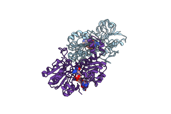 |
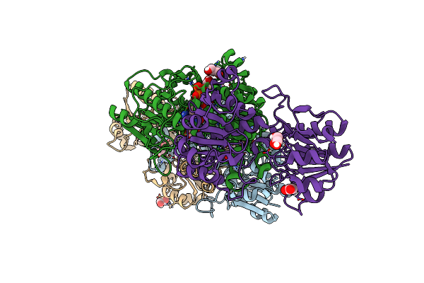
Formate Dehydrogenase From Novosphingobium Sp. Ap12 With Nadp And Azide
Organism: Novosphingobium sp. ap12
Method: X-RAY DIFFRACTION Resolution:3.00 Å Release Date: 2023-07-12 Classification: OXIDOREDUCTASE Ligands: NAP, AZI |
Organism: Novosphingobium sp. ap12
Method: X-RAY DIFFRACTION
Release Date: 2023-07-12
Ligands: NAP, AZI
 |
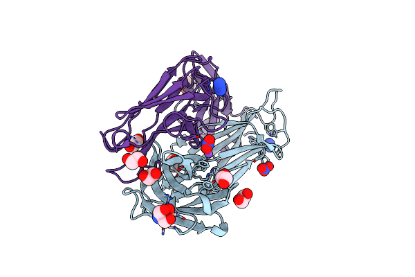
Formate Dehydrogenase From Novosphingobium Sp. Ap12 With Nad And Azide
Organism: Novosphingobium sp. ap12
Method: X-RAY DIFFRACTION Resolution:1.80 Å Release Date: 2023-07-12 Classification: OXIDOREDUCTASE Ligands: NAD, AZI, EDO, NA, GOL, ACT |
Organism: Novosphingobium sp. ap12
Method: X-RAY DIFFRACTION
Release Date: 2023-07-12
Ligands: NAD, AZI, EDO, NA, GOL, ACT
 |
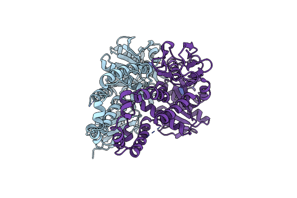
Crystal Structure Of The Human Igd Fab - Structure Fab2
Organism: Homo sapiens
Method: X-RAY DIFFRACTION Resolution:1.55 Å Release Date: 2023-06-14 Classification: IMMUNE SYSTEM Ligands: NO3, AZI, EDO, NA, TRS |
Organism: Homo sapiens
Method: X-RAY DIFFRACTION
Release Date: 2023-06-14
Ligands: NO3, AZI, EDO, NA, TRS
 |
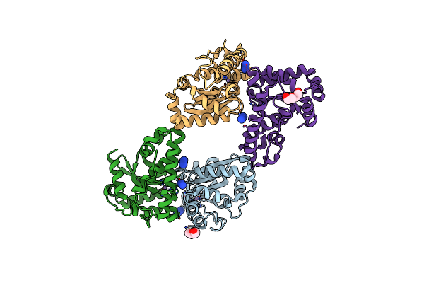
Re-Refinement Of Crystal Structure Of Nosget3D, The All4481 Protein From Nostoc Sp. Pcc 7120
Organism: Nostoc sp. pcc 7120 = fachb-418
Method: X-RAY DIFFRACTION Resolution:1.98 Å Release Date: 2023-05-24 Classification: CHAPERONE Ligands: AZI |
Organism: Nostoc sp. pcc 7120 = fachb-418
Method: X-RAY DIFFRACTION
Release Date: 2023-05-24
Ligands: AZI
 |
Crystal Structure Of A Mutant Staphylococcus Equorum Manganese Superoxide Dismutase K38R And A121E
Organism: Staphylococcus equorum
Method: X-RAY DIFFRACTION Resolution:1.40 Å Release Date: 2023-01-25 Classification: OXIDOREDUCTASE Ligands: MN, PEG, AZI, BR |
Organism: Staphylococcus equorum
Method: X-RAY DIFFRACTION
Release Date: 2023-01-25
Ligands: MN, PEG, AZI, BR
 |
Crystal Structure Of A Mutant Staphylococcus Equorum Manganese Superoxide Dismutase K38R And A121Y
Organism: Staphylococcus equorum
Method: X-RAY DIFFRACTION Resolution:1.51 Å Release Date: 2023-01-25 Classification: OXIDOREDUCTASE Ligands: PEG, MN, BR, TRS, AZI |
Organism: Staphylococcus equorum
Method: X-RAY DIFFRACTION
Release Date: 2023-01-25
Ligands: PEG, MN, BR, TRS, AZI
 |
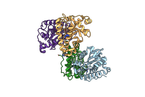
Crystal Structure Of Formate Dehydrogenase Fdh2 Enzyme From Granulicella Mallensis Mp5Actx8 In Complex With Nad And Azide.
Organism: Granulicella mallensis mp5actx8
Method: X-RAY DIFFRACTION Resolution:1.97 Å Release Date: 2023-01-18 Classification: OXIDOREDUCTASE Ligands: NAD, AZI, EDO |
Organism: Granulicella mallensis mp5actx8
Method: X-RAY DIFFRACTION
Release Date: 2023-01-18
Ligands: NAD, AZI, EDO
 |
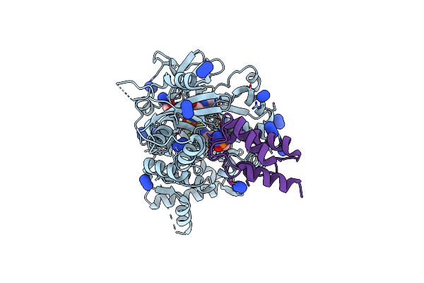
Crystal Structure Of A Mutant Staphylococcus Equorum Manganese Superoxide Dismutase L169W
Organism: Staphylococcus equorum
Method: X-RAY DIFFRACTION Resolution:1.94 Å Release Date: 2022-12-07 Classification: OXIDOREDUCTASE Ligands: MN, AZI |
Organism: Staphylococcus equorum
Method: X-RAY DIFFRACTION
Release Date: 2022-12-07
Ligands: MN, AZI
 |
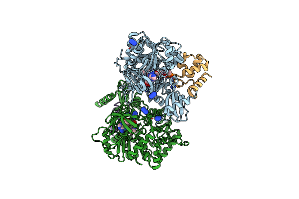
Crystal Structure Of Pigi Trapped With Pigg Using A Proline Adenosine Vinylsulfonamide Inhibitor
Organism: Serratia sp. atcc 39006
Method: X-RAY DIFFRACTION Resolution:1.60 Å Release Date: 2022-11-30 Classification: LIGASE Ligands: GOL, AZI, MG, I5M |
Organism: Serratia sp. atcc 39006
Method: X-RAY DIFFRACTION
Release Date: 2022-11-30
Ligands: GOL, AZI, MG, I5M
 |
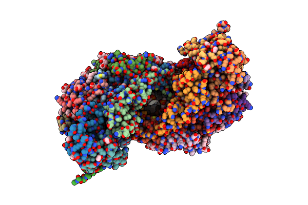
Crystal Structure Of Pltf Trapped With Pigg Using A Proline Adenosine Vinylsulfonamide Inhibitor
Organism: Pseudomonas protegens pf-5, Serratia sp. atcc 39006
Method: X-RAY DIFFRACTION Resolution:2.46 Å Release Date: 2022-11-30 Classification: LIGASE/LIGASE INHIBITOR Ligands: AZI, I5M |
Organism: Pseudomonas protegens pf-5, Serratia sp. atcc 39006
Method: X-RAY DIFFRACTION
Release Date: 2022-11-30
Ligands: AZI, I5M
 |
The Structure Of Azide-Bound Cytochrome C Oxidase Determined Using The Crystals Exposed To 20 Mm Azide Solution For 4 Days
Organism: Bos taurus
Method: X-RAY DIFFRACTION Resolution:1.85 Å Release Date: 2022-10-19 Classification: OXIDOREDUCTASE Ligands: HEA, CU, MG, NA, AZI, PGV, EDO, TGL, CHD, CUA, PSC, DMU, CDL, PEK, ZN |