
Search Count: 31
All
Selected
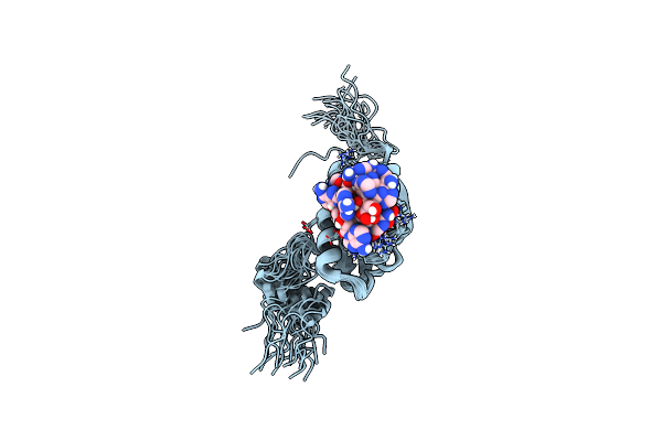 |
Organism: Rhodobacter sphaeroides 2.4.1
Method: SOLUTION NMR Release Date: 2005-12-07 Classification: APPA Ligands: FAD |
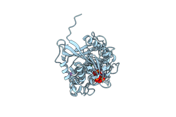 |
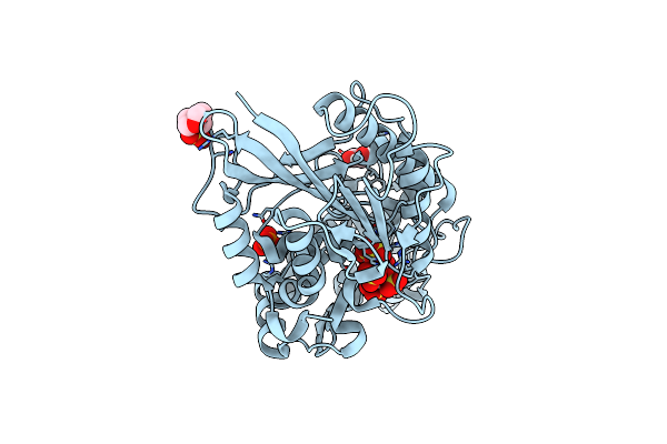
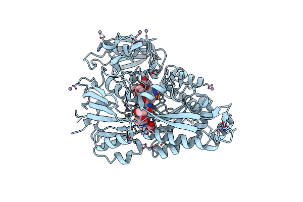
Escherichia Coli Periplasmic Phytase Appa, Complex With Myo-Inositol Hexakissulfate
Organism: Escherichia coli
Method: X-RAY DIFFRACTION Resolution:1.72 Å Release Date: 2022-03-16 Classification: HYDROLASE Ligands: IHS, NI, K |
 |
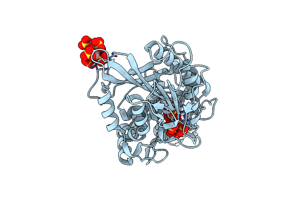
Organism: Escherichia coli k-12
Method: X-RAY DIFFRACTION Resolution:1.85 Å Release Date: 2022-03-16 Classification: HYDROLASE Ligands: PO4, NI, MG |
 |
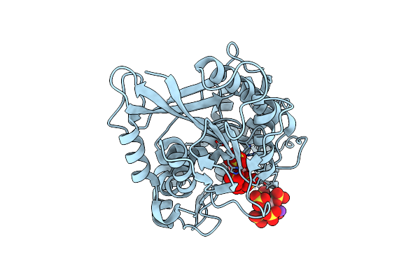
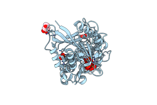
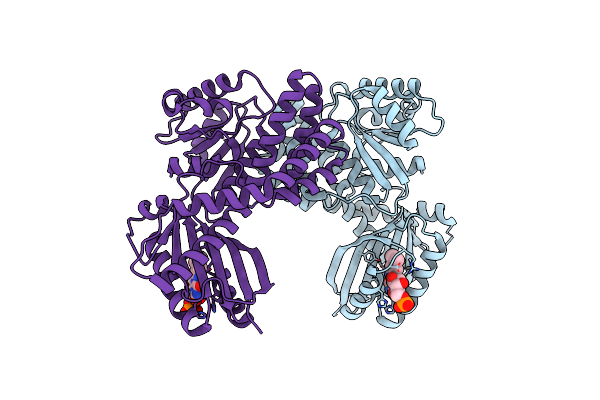
Escherichia Coli Periplasmic Phytase Appa T305E Mutant, Complex With Myo-Inositol Hexakissulfate
Organism: Escherichia coli
Method: X-RAY DIFFRACTION Resolution:1.86 Å Release Date: 2022-03-16 Classification: HYDROLASE Ligands: IHS, NI |
 |
Escherichia Coli Periplasmic Phytase Appa D304E Mutant, Complex With Myo-Inositol Hexakissulfate
Organism: Escherichia coli
Method: X-RAY DIFFRACTION Resolution:2.60 Å Release Date: 2022-06-29 Classification: HYDROLASE Ligands: IHS, K |
 |
Escherichia Coli Periplasmic Phytase Appa D304A Mutant, Complex With Myo-Inositol Hexakissulfate
Organism: Escherichia coli
Method: X-RAY DIFFRACTION Resolution:1.41 Å Release Date: 2022-03-16 Classification: HYDROLASE Ligands: IHS, GOL, PO4, MES |
 |
Escherichia Coli Periplasmic Phytase Appa D304A,T305E Mutant, Complex With Myo-Inositol Hexakissulfate
Organism: Escherichia coli
Method: X-RAY DIFFRACTION Resolution:1.42 Å Release Date: 2022-03-16 Classification: HYDROLASE Ligands: IHS, GOL, PO4, MES |
 |
Escherichia Coli Periplasmic Phytase Appa D304A Mutant, Phosphohistidine Intermediate
Organism: Escherichia coli
Method: X-RAY DIFFRACTION Resolution:1.85 Å Release Date: 2022-03-16 Classification: HYDROLASE Ligands: NI |
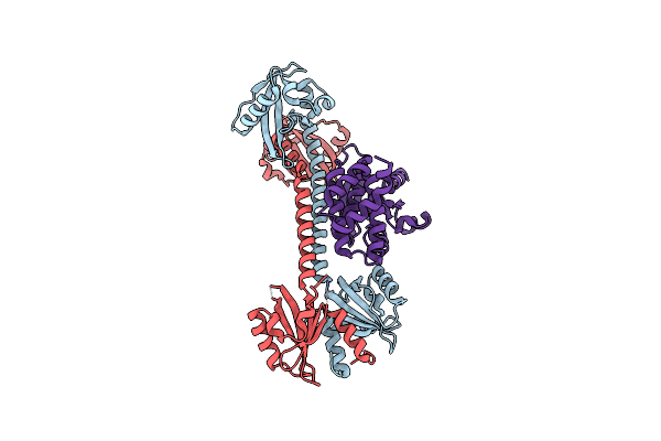 |
Organism: Rhodobacter sphaeroides
Method: X-RAY DIFFRACTION Resolution:1.75 Å Release Date: 2013-06-05 Classification: FLAVOPROTEIN/TRANSCRIPTION |
 |
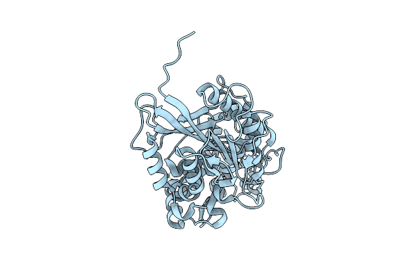
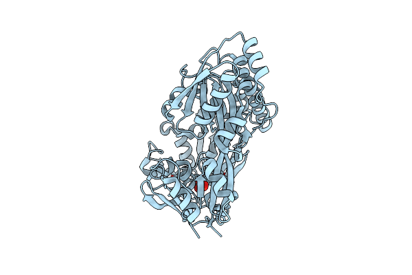
The Structure Of The Oligopeptide-Binding Protein, Appa, From Bacillus Subtilis In Complex With A Nonapeptide.
Organism: Bacillus subtilis
Method: X-RAY DIFFRACTION Resolution:1.55 Å Release Date: 2005-01-25 Classification: TRANSPORT PROTEIN Ligands: ZN |
 |
Crystal Structure Of A Putative Peptide Binding Protein Appa From Clostridium Difficile
Organism: Clostridioides difficile
Method: X-RAY DIFFRACTION Resolution:2.00 Å Release Date: 2019-04-10 Classification: PEPTIDE BINDING PROTEIN Ligands: IOD, SO4 |
 |
Organism: Rhodobacter sphaeroides
Method: X-RAY DIFFRACTION Resolution:2.05 Å Release Date: 2013-09-18 Classification: METAL BINDING PROTEIN |
 |
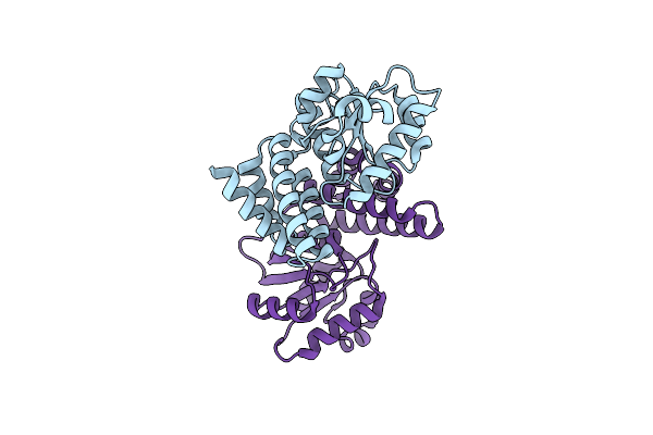
Dark-State Structure Of Appa C20S Without The Cys-Rich Region From Rb. Sphaeroides
Organism: Rhodobacter sphaeroides
Method: X-RAY DIFFRACTION Resolution:2.60 Å Release Date: 2013-06-05 Classification: FLAVOPROTEIN,SIGNALING PROTEIN Ligands: FMN, CL |
 |
Dark-State Structure Of Appa Wild-Type Without The Cys-Rich Region From Rb. Sphaeroides
Organism: Rhodobacter sphaeroides
Method: X-RAY DIFFRACTION Resolution:3.50 Å Release Date: 2013-06-05 Classification: FLAVOPROTEIN,SIGNALING PROTEIN Ligands: FMN |
 |
Structure Of A Novel Photoreceptor: The Bluf Domain Of Appa From Rhodobacter Sphaeroides
Organism: Rhodobacter sphaeroides 2.4.1
Method: X-RAY DIFFRACTION Resolution:2.30 Å Release Date: 2005-06-28 Classification: TRANSCRIPTION Ligands: FMN, D9G |
 |
NA
Organism: Escherichia coli (strain K12)
Method: Alphafold Release Date: Classification: NA Ligands: NA |
 |
NA
Organism: Bacillus subtilis (strain 168)
Method: Alphafold Release Date: Classification: NA Ligands: NA |
 |
Organism: Nicotiana tabacum
Method: X-RAY DIFFRACTION Resolution:3.20 Å Release Date: 2001-09-05 Classification: LYASE Ligands: HEN |
 |
Organism: Rhodobacter sphaeroides
Method: X-RAY DIFFRACTION Resolution:2.30 Å Release Date: 2006-09-06 Classification: SIGNAL TRANSDUCTION Ligands: FMN, DTU, DTT |
 |
Structure Of A Light-Induced Intermediate Of The Bluf Domain Of The Rhodobacterial Protein Appa
Organism: Rhodobacter sphaeroides
Method: X-RAY DIFFRACTION Resolution:2.95 Å Release Date: 2006-09-06 Classification: SIGNAL TRANSDUCTION Ligands: FMN, DTU, DTT |