
Search Count: 3,930
 |
Organism: Pseudomonas aeruginosa pao1
Method: X-RAY DIFFRACTION Resolution:2.25 Å Release Date: 2020-12-02 Classification: ANTIBIOTIC Ligands: UQ8 |
 |
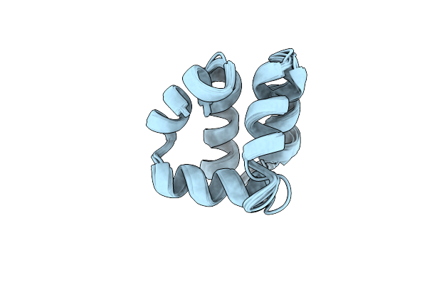
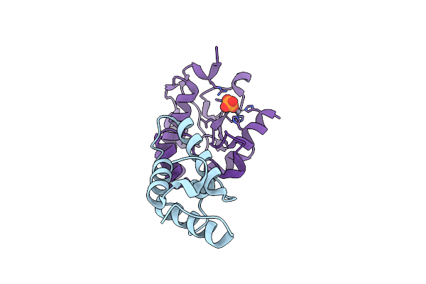
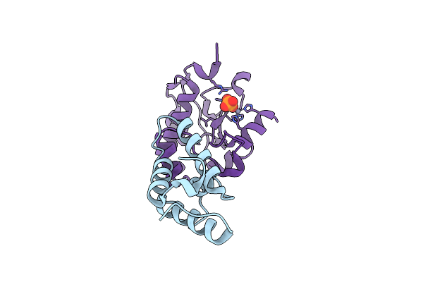
Organism: Streptomyces globisporus
Method: X-RAY DIFFRACTION Resolution:1.80 Å Release Date: 2003-06-03 Classification: ANTIBIOTIC |
 |
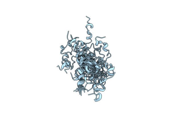
An Antibiotic Biosynthesis Monooxygenase Family Protein From Streptomyces Sp. Ma37
Organism: Streptomyces sp. ma37
Method: X-RAY DIFFRACTION Resolution:1.65 Å Release Date: 2025-11-26 Classification: ANTIBIOTIC |
 |
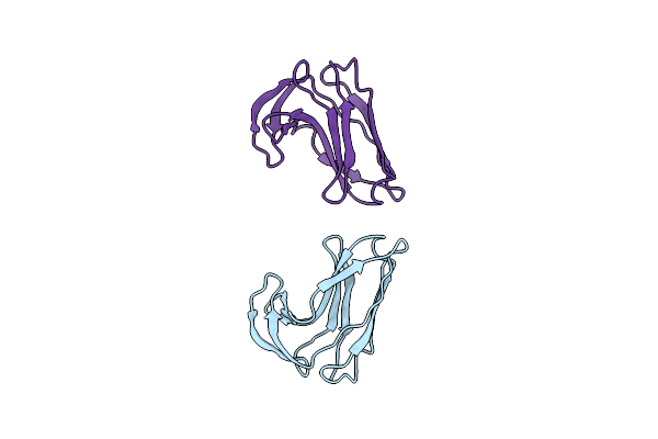
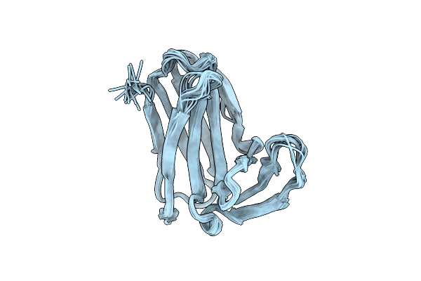
Solution Structures Of C-1027 Apoprotein And Its Complex With The Aromatized Chromophore
Organism: Streptomyces globisporus
Method: SOLUTION NMR Release Date: 2001-05-23 Classification: ANTIBIOTIC |
 |
Solution Structures Of C-1027 Apoprotein And Its Complex With The Aromatized Chromophore
Organism: Streptomyces globisporus
Method: SOLUTION NMR Release Date: 2001-05-23 Classification: ANTIBIOTIC Ligands: ROM |
 |
Organism: Enterococcus faecalis
Method: SOLUTION NMR Release Date: 2000-10-25 Classification: ANTIBIOTIC |
 |
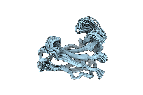
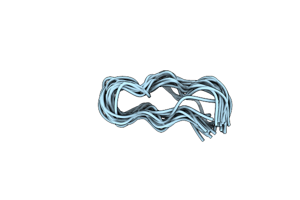
Sequential 1H,13C And 15N Nmr Assignments And Solution Conformation Of Apokedarcidin
Organism: Actinomycete atcc 53650
Method: SOLUTION NMR Release Date: 1994-08-31 Classification: ANTIBIOTIC CHROMOPROTEIN |
 |
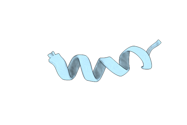
Solution Structure Of A Beta-Hairpin Peptidomimetic Antibiotic That Target Lptd In Pseudomonas Sp.
|
 |
Organism: Enterococcus faecalis
Method: X-RAY DIFFRACTION Resolution:1.20 Å Release Date: 2015-04-15 Classification: ANTIBIOTIC Ligands: CIT |
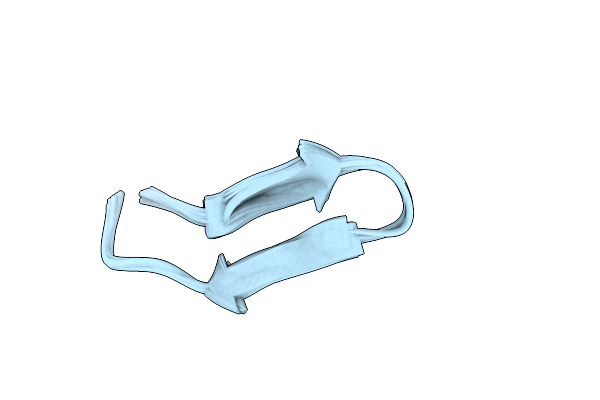 |
Solution Structure Of A Beta-Hairpin Peptidomimetic Antibiotic That Targets Lptd In Pseudomonas Sp.
|
 |
Organism: Nocardiopsis alba (strain atcc baa-2165 / be74)
Method: SOLUTION NMR Release Date: 2017-03-01 Classification: ANTIBIOTIC |
 |
Nmr Structure Of Zervamicin Iib (Peptaibol Antibiotic) Bound To Dpc Micelles
Organism: Emericellopsis salmosynnemata
Method: SOLUTION NMR Release Date: 2002-02-13 Classification: ANTIBIOTIC |
 |
 |
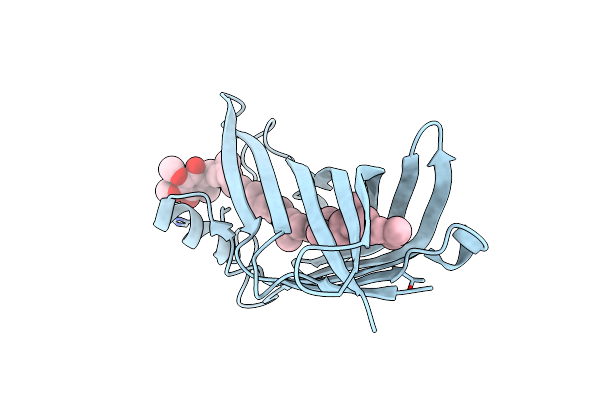
Organism: Klebsiella pneumoniae
Method: X-RAY DIFFRACTION Resolution:1.50 Å Release Date: 2019-10-09 Classification: HYDROLASE/Antibiotic/Inhibitor Ligands: 5R7, EDO, CL, EPE |
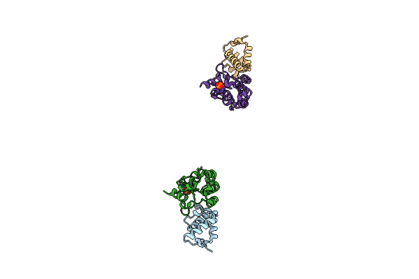 |
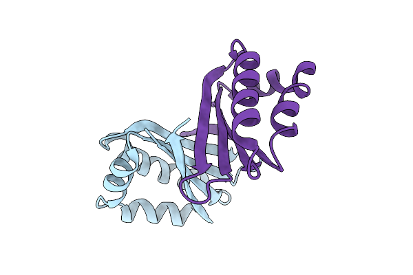
Crystal Structure Of The E9 Dnase Domain With A Mutant Immunity Protein Im9 (D51A)
Organism: Escherichia coli k12
Method: X-RAY DIFFRACTION Resolution:1.60 Å Release Date: 2008-03-18 Classification: antibiotic/antibiotic inhibitor Ligands: ZN, PO4 |
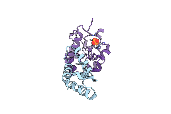 |
Crystal Structure Of The E9 Dnase Domain With A Mutant Immunity Protein Im9 (Y54F)
Organism: Escherichia coli k12
Method: X-RAY DIFFRACTION Resolution:1.75 Å Release Date: 2007-07-03 Classification: Antibiotic/Antibiotic Inhibitor Ligands: ZN, PO4 |
 |
Crystal Structure Of The E9 Dnase Domain With A Mutant Immunity Protein Im9 (V34A)
Organism: Escherichia coli k12
Method: X-RAY DIFFRACTION Resolution:1.70 Å Release Date: 2008-03-18 Classification: antibiotic/antibiotic inhibitor Ligands: ZN, PO4 |
 |
Crystal Structure Of The Complex Of The Colicin E9 Dnase Domain With A Mutant Immunity Protein, Imme9 (D51A)
Organism: Escherichia coli k12
Method: X-RAY DIFFRACTION Resolution:1.60 Å Release Date: 2007-05-15 Classification: antibiotic/antibiotic inhibitor Ligands: ZN, PO4 |
 |
Crystal Structure Of The E9 Dnase Domain With A Mutant Immunity Protein Im9 (Y55A)
Organism: Escherichia coli k12
Method: X-RAY DIFFRACTION Resolution:1.80 Å Release Date: 2008-03-18 Classification: Antibiotic/Antibiotic Inhibitor Ligands: ZN, PO4 |
 |
Crystal Structure Of The E9 Dnase Domain With A Mutant Immunity Protein Im9 (Y55F)
Organism: Escherichia coli k12
Method: X-RAY DIFFRACTION Resolution:1.70 Å Release Date: 2008-03-18 Classification: antibiotic/antibiotic inhibitor Ligands: ZN, PO4 |