
Search Count: 787
 |
Organism: Homo sapiens
Method: X-RAY DIFFRACTION Resolution:1.85 Å Release Date: 2025-09-24 Classification: HYDROLASE Ligands: MG, SO4, ACY, ADP |
 |
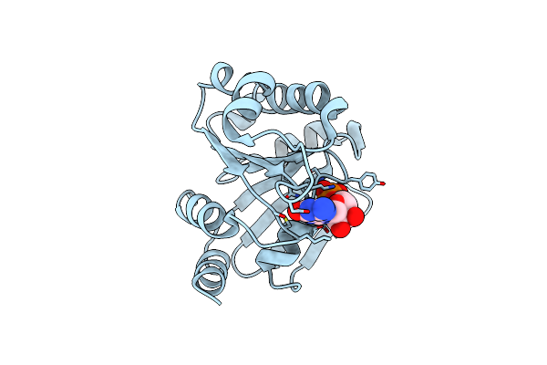
Adp-Ribosylserine Hydrolase Arh3 Of Latimeria Chalumnae In Complex With Adp-Ribosyl-L-Arginine
Organism: Latimeria chalumnae
Method: X-RAY DIFFRACTION Resolution:1.66 Å Release Date: 2018-11-28 Classification: HYDROLASE Ligands: MG, F2R, GOL, ACT |
 |
Human Adp-Ribosylserine Hydrolase Arh3 Mutant E41A In Complex With H2B-S7-Mar Peptide
Organism: Homo sapiens
Method: X-RAY DIFFRACTION Resolution:1.86 Å Release Date: 2021-06-16 Classification: HYDROLASE Ligands: MG, EDO, ACT, AR6 |
 |
Human Adp-Ribosylserine Hydrolase Arh3 Mutant E41A In Complex With Adp-Ribose Dimer
Organism: Homo sapiens
Method: X-RAY DIFFRACTION Resolution:1.95 Å Release Date: 2021-06-16 Classification: HYDROLASE Ligands: MG, AR6, CL, EDO |
 |
Organism: Homo sapiens
Method: X-RAY DIFFRACTION Resolution:1.80 Å Release Date: 2018-11-28 Classification: HYDROLASE Ligands: A3R, MG, CL, ACT |
 |
Organism: Homo sapiens
Method: X-RAY DIFFRACTION Resolution:1.85 Å Release Date: 2025-09-24 Classification: HYDROLASE Ligands: MG, ACY, A1JFJ |
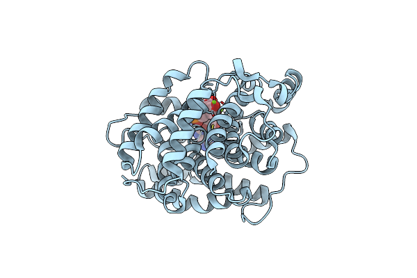 |
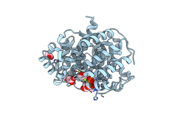
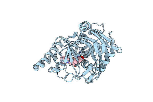
Human [Protein Adp-Ribosylargenine] Hydrolase Arh1 In Complex With Adp-Ribose
Organism: Homo sapiens
Method: X-RAY DIFFRACTION Resolution:1.23 Å Release Date: 2018-11-28 Classification: HYDROLASE Ligands: AR6, MG, CL |
 |
Organism: Homo sapiens
Method: X-RAY DIFFRACTION Resolution:2.96 Å Release Date: 2020-02-19 Classification: TRANSFERASE Ligands: UHB |
 |
Organism: Leishmania major
Method: X-RAY DIFFRACTION Resolution:2.10 Å Release Date: 2010-03-02 Classification: GTP-BINDING PROTEIN Ligands: GDP, MG |
 |
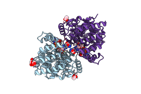
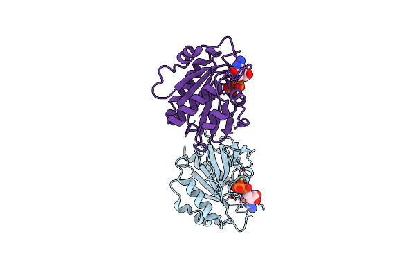
Structure Of An Adp Ribosylation Factor From Entamoeba Histolytica Hm-1:Imss Bound To Mg-Gdp
Organism: Entamoeba histolytica
Method: X-RAY DIFFRACTION Resolution:1.80 Å Release Date: 2015-03-04 Classification: SIGNALING PROTEIN Ligands: GDP, MG |
 |
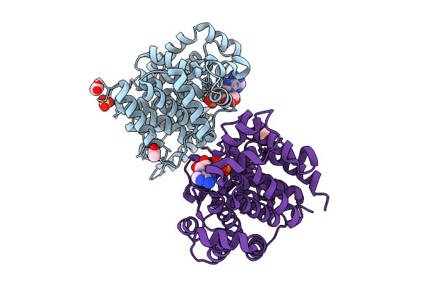
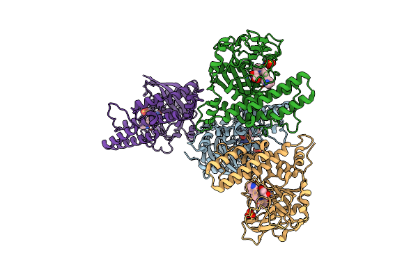
Organism: Homo sapiens, Bos taurus
Method: X-RAY DIFFRACTION Resolution:3.30 Å Release Date: 2013-09-25 Classification: PROTEIN TRANSPORT Ligands: G3D |
 |
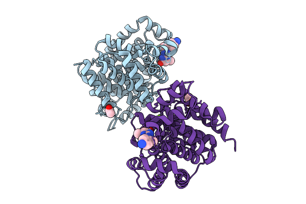
Domain Iii Of Pseudomonas Aeruginosa Exotoxin Complexed With Nicotinamide And Amp
Organism: Pseudomonas aeruginosa
Method: X-RAY DIFFRACTION Resolution:2.50 Å Release Date: 1995-09-15 Classification: ADP-RIBOSYLATION Ligands: NCA, AMP |
 |
Organism: Homo sapiens
Method: X-RAY DIFFRACTION Resolution:3.19 Å Release Date: 2015-12-09 Classification: Transferase/Transferase inhibitor Ligands: SO4, RPB |
 |
Organism: Homo sapiens
Method: X-RAY DIFFRACTION Resolution:1.90 Å Release Date: 2006-06-13 Classification: TRANSPORT PROTEIN Ligands: GDP, UNX |
 |
Organism: Homo sapiens
Method: X-RAY DIFFRACTION Resolution:2.68 Å Release Date: 2014-01-01 Classification: TRANSPORT PROTEIN Ligands: GDP |
 |
Organism: Homo sapiens
Method: X-RAY DIFFRACTION Resolution:2.84 Å Release Date: 2015-09-02 Classification: TRANSFERASE/TRANSFERASE INHIBITOR Ligands: SO4, XAV |
 |
Organism: Homo sapiens
Method: X-RAY DIFFRACTION Resolution:2.20 Å Release Date: 2015-09-09 Classification: TRANSFERASE/TRANSFERASE INHIBITOR Ligands: 3JD, SO4, GOL |
 |
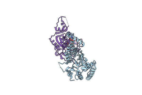
The Catalytic Fragment Of Poly(Adp-Ribose) Polymerase Complexed With An Isoindolinone Inhibitor
Organism: Gallus gallus
Method: X-RAY DIFFRACTION Resolution:2.10 Å Release Date: 2019-05-01 Classification: TRANSFERASE Ligands: H7Z |
 |
The Catalytic Fragment Of Poly(Adp-Ribose) Polymerase Complexed With Isoindolinone Inhibitor
Organism: Gallus gallus
Method: X-RAY DIFFRACTION Resolution:2.10 Å Release Date: 2019-05-01 Classification: TRANSFERASE Ligands: H7W |
 |
Organism: Pseudomonas aeruginosa
Method: X-RAY DIFFRACTION Resolution:2.30 Å Release Date: 1996-06-10 Classification: ADP-RIBOSYLATION Ligands: TAD, TIA, AMP |