
Search Count: 4
All
Selected
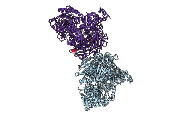 |
Crystal Structure Of N-Methylhydantoinase In Complex With 1-Methylimidazolidine-2,4-Dione
Organism: Glutamicibacter protophormiae
Method: X-RAY DIFFRACTION Resolution:2.07 Å Release Date: 2026-04-15 Classification: HYDROLASE Ligands: CA, A1BC1, NH4, BTB |
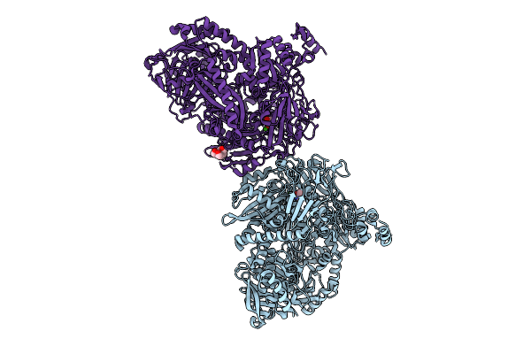 |
Crystal Structure Of N-Methylhydantoinase In Complex With 1-Methylimidazolidine-2,4-Dione, Iodide Soak
Organism: Glutamicibacter protophormiae
Method: X-RAY DIFFRACTION Resolution:2.62 Å Release Date: 2026-04-15 Classification: HYDROLASE Ligands: CA, A1BC1, NH4, IOD, BTB |
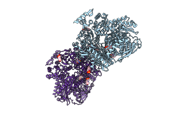 |
Crystal Structure Of N-Methylhydantoinase In Complex With 1-Methylimidazolidine-2,4-Dione, C2221 Form
Organism: Glutamicibacter protophormiae
Method: X-RAY DIFFRACTION Resolution:2.80 Å Release Date: 2026-04-15 Classification: HYDROLASE Ligands: CA, A1BC1, NH4, MES, SO4 |
 |
Crystal Structure Of N-Methylhydantoinase In Complex With 1-Methylimidazolidine-2,4-Dione, C-Terminal Residues Visible
Organism: Glutamicibacter protophormiae
Method: X-RAY DIFFRACTION Resolution:2.07 Å Release Date: 2026-04-15 Classification: HYDROLASE Ligands: CA, A1BC1, BTB, NH4, SO4 |