
Search Count: 1,420
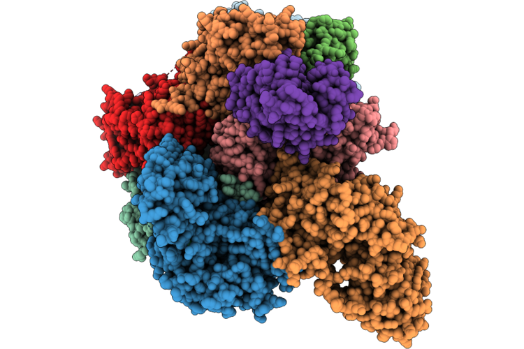 |
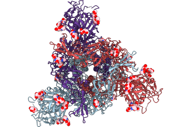
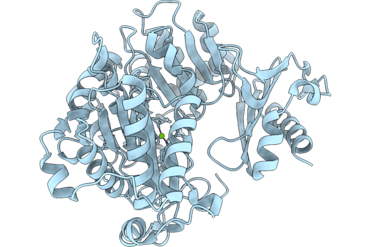
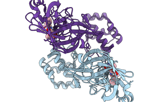
Eukaryotic Translation Initiation Factor 2-B (Eif2B) Bound To The Viral Effector Acp10
Organism: Homo sapiens, Beluga whale coronavirus sw1
Method: ELECTRON MICROSCOPY Release Date: 2026-06-24 Classification: TRANSLATION Ligands: CL, MG, ZN |
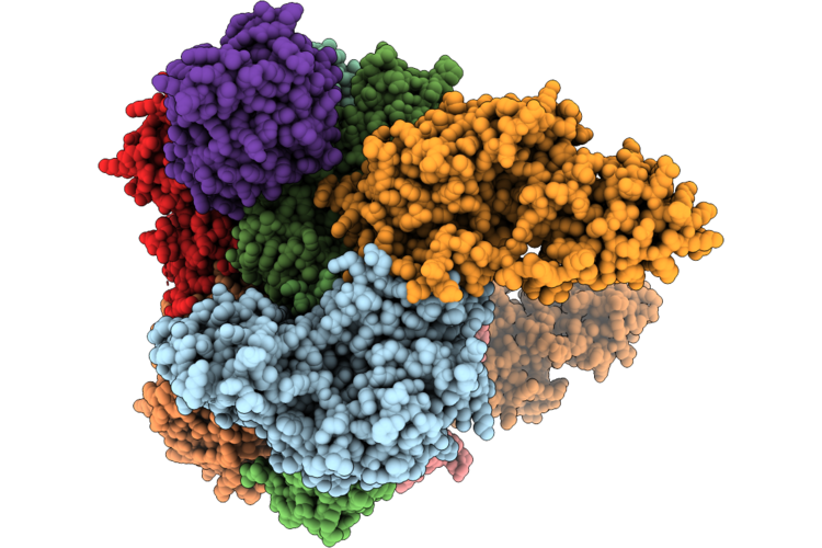 |
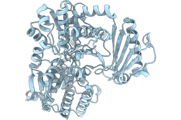
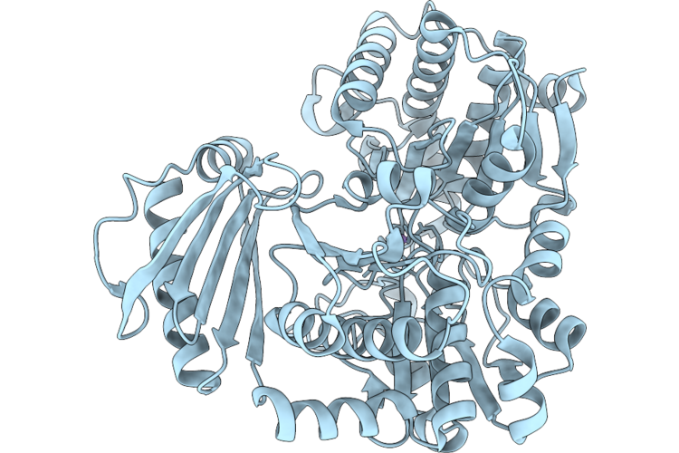
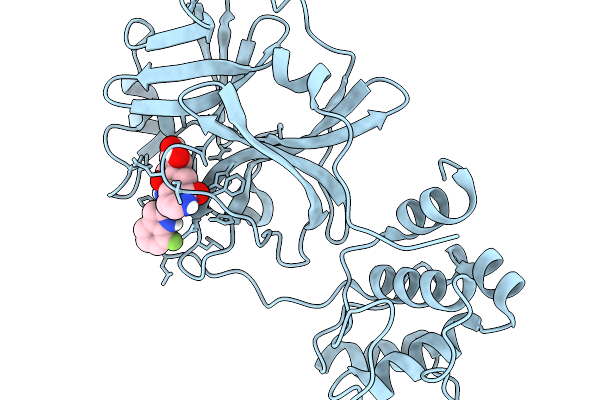
Eukaryotic Translation Initiation Factor 2-B (Eif2B) With A Truncation In The Beta Subunit (Active-Like-State) Bound To The Viral Effector Acp10
Organism: Homo sapiens, Beluga whale coronavirus sw1
Method: ELECTRON MICROSCOPY Resolution:2.10 Å Release Date: 2026-06-24 Classification: TRANSLATION Ligands: CL, PO4, ZN |
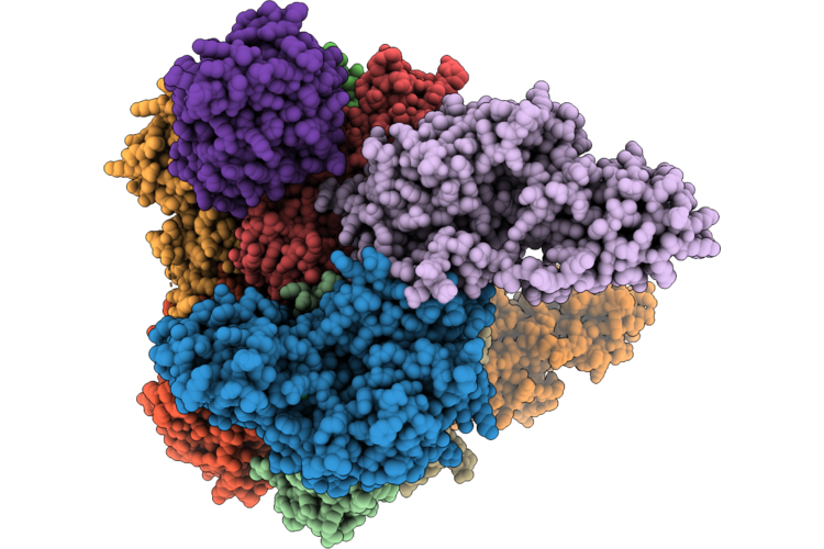 |
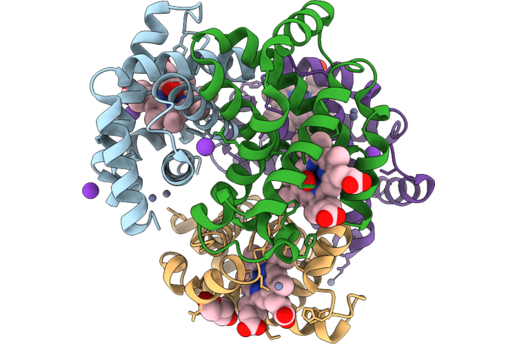
Eukaryotic Translation Initiation Factor 2-B (Eif2B) With A Truncation In The Beta Subunit (Active-Like-State) Asymmetrically Bound To The Viral Effector Acp10
Organism: Homo sapiens, Beluga whale coronavirus sw1
Method: ELECTRON MICROSCOPY Release Date: 2026-06-24 Classification: TRANSLATION Ligands: CL, PO4, ZN |
 |
Organism: Promethearchaeum syntrophicum
Method: X-RAY DIFFRACTION Resolution:1.90 Å Release Date: 2026-06-17 Classification: LYASE Ligands: PO4 |
 |
Organism: Phaeodactylibacter sp.
Method: X-RAY DIFFRACTION Resolution:1.58 Å Release Date: 2026-06-10 Classification: ISOMERASE Ligands: MG |
 |
Organism: Bat coronavirus hku4
Method: ELECTRON MICROSCOPY Release Date: 2026-05-27 Classification: VIRAL PROTEIN Ligands: NAG |
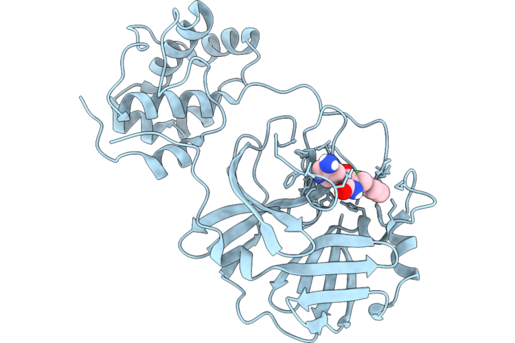 |
The Crystal Structure Of Sars-Cov-1 Main Protease In Complex With Inhibitor Fd2-21
Organism: Severe acute respiratory syndrome-related coronavirus
Method: X-RAY DIFFRACTION Resolution:1.73 Å Release Date: 2026-05-27 Classification: VIRAL PROTEIN Ligands: A1EZ7 |
 |
Crystal Structure Of Phaeodactylibacter Sp. Phosphoglucomutase In Complex With Glucose-1-Phosphate
Organism: Phaeodactylibacter sp.
Method: X-RAY DIFFRACTION Resolution:1.95 Å Release Date: 2026-05-13 Classification: ISOMERASE Ligands: MG, G1P |
 |
Crystal Structure Of Phaeodactylibacter Sp. Phosphoglucomutase In Complex With Magnesium Ion
Organism: Phaeodactylibacter sp.
Method: X-RAY DIFFRACTION Resolution:1.65 Å Release Date: 2026-05-13 Classification: ISOMERASE Ligands: MG |
 |
Crystal Structure Of Phaeodactylibacter Sp. Phosphoglucomutase In Complex With Manganese Ion
Organism: Phaeodactylibacter sp.
Method: X-RAY DIFFRACTION Resolution:1.82 Å Release Date: 2026-05-13 Classification: ISOMERASE Ligands: MN |
 |
Crystal Structure Of Sheep (Ovis Aries) Oxyhemoglobin At 2.1 Angstrom Resolution
Organism: Ovis aries
Method: X-RAY DIFFRACTION Resolution:2.08 Å Release Date: 2026-05-06 Classification: OXYGEN TRANSPORT Ligands: HEM, OXY, K, ZN, PEG |
 |
Crystal Structure Of Phaeodactylibacter Sp. Phosphoglucomutase In Complex With Glucose-6-Phosphate
Organism: Phaeodactylibacter sp.
Method: X-RAY DIFFRACTION Resolution:1.97 Å Release Date: 2026-04-15 Classification: ISOMERASE Ligands: G6P, MG |
 |
Organism: Severe acute respiratory syndrome-related coronavirus
Method: X-RAY DIFFRACTION Resolution:2.04 Å Release Date: 2026-03-11 Classification: VIRAL PROTEIN Ligands: YDL |
 |
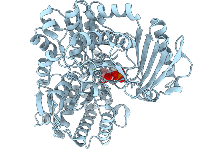
Crystal Structure Of Sars Main Protease In Complex With Ibuzatrelvir Pomotrelvir
Organism: Severe acute respiratory syndrome-related coronavirus
Method: X-RAY DIFFRACTION Resolution:2.05 Å Release Date: 2026-03-04 Classification: VIRAL PROTEIN Ligands: ZQB |
 |
Crystal Structure Of Monomeric Rag-Like Small Gtpase From Asgard Lokiarchaeota (Lokiragm) In Complex With Gtp
Organism: Promethearchaeum syntrophicum
Method: X-RAY DIFFRACTION Resolution:2.00 Å Release Date: 2026-03-04 Classification: HYDROLASE Ligands: EDO, GOL, MG, GTP |
 |
Organism: Tylonycteris bat coronavirus hku4
Method: X-RAY DIFFRACTION Resolution:2.05 Å Release Date: 2026-02-18 Classification: VIRUS Ligands: A1BWH |
 |
Organism: Miniopterus bat coronavirus hku8
Method: X-RAY DIFFRACTION Resolution:1.80 Å Release Date: 2026-02-18 Classification: VIRAL PROTEIN Ligands: A1BWH |
 |
Crystal Structure Of The Daba Transaminase Ectb From The Halophilic And Cold-Adapted Marinobacter Sp. Ck1 -Mutant K264A
Organism: Marinobacter sp. ck-1
Method: X-RAY DIFFRACTION Resolution:1.55 Å Release Date: 2026-02-18 Classification: TRANSFERASE Ligands: PLP, EDO, SO4, NA |
 |
Crystal Structure Of The Daba Transaminase Ectb From The Halophilic And Cold-Adapted Marinobacter Sp. Ck1
Organism: Marinobacter sp. ck-1
Method: X-RAY DIFFRACTION Resolution:2.00 Å Release Date: 2026-02-18 Classification: TRANSFERASE Ligands: PMP, GOL |
 |
Organism: Beluga whale coronavirus sw1
Method: ELECTRON MICROSCOPY Resolution:2.24 Å Release Date: 2026-02-11 Classification: VIRAL PROTEIN Ligands: NAG |