
Search Count: 957
 |
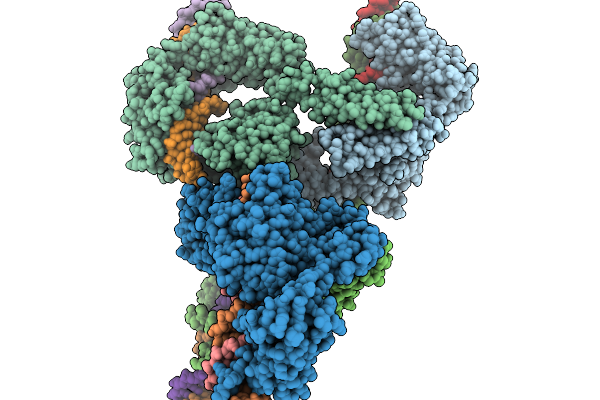
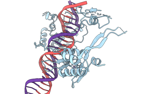
Cryo-Em Structure Of The Large Serine Recombinase Bxb1 In Complex With Attp And Attb (Gt/Tt Cdn) In The Pre-Strand Exchange State
Organism: Mycobacterium phage bxb1, Mycolicibacterium smegmatis
Method: ELECTRON MICROSCOPY Resolution:3.22 Å Release Date: 2026-03-04 Classification: DNA BINDING PROTEIN Ligands: ZN |
 |
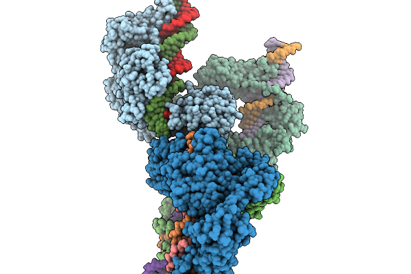
Cryo-Em Structure Of The Large Serine Recombinase Bxb1 In Complex With Attp And Attb (Gt/Tt Cdn) In The Post-Strand Exchange State
Organism: Mycobacterium phage bxb1, Mycolicibacterium smegmatis
Method: ELECTRON MICROSCOPY Resolution:3.97 Å Release Date: 2026-03-04 Classification: DNA BINDING PROTEIN Ligands: ZN |
 |
Cryo-Em Structure Of The Large Serine Recombinase Bxb1 In Complex With Attp And Attb (Ca/Ca Cdn) In The Pre-Strand Exchange State
Organism: Mycobacterium phage bxb1, Mycolicibacterium smegmatis
Method: ELECTRON MICROSCOPY Resolution:3.58 Å Release Date: 2026-03-04 Classification: DNA BINDING PROTEIN Ligands: ZN |
 |
Cryo-Em Structure Of The Large Serine Recombinase Bxb1 In Complex With Attp And Attb (Ca/Ca Cdn) In The Post-Strand Exchange State
Organism: Mycobacterium phage bxb1, Mycolicibacterium smegmatis
Method: ELECTRON MICROSCOPY Resolution:3.63 Å Release Date: 2026-03-04 Classification: DNA BINDING PROTEIN Ligands: ZN |
 |
Cryo-Em Structure Of The Large Serine Recombinase Bxb1 In Complex With Attp And Attb (Ca/Ca Cdn) In The Intermediate-Strand Exchange State 1
Organism: Mycobacterium phage bxb1, Mycolicibacterium smegmatis
Method: ELECTRON MICROSCOPY Resolution:3.97 Å Release Date: 2026-03-04 Classification: DNA BINDING PROTEIN Ligands: ZN |
 |
Cryo-Em Structure Of The Large Serine Recombinase Bxb1 In Complex With Attp And Attb (Ca/Ca Cdn) In The Intermediate-Strand Exchange State 2
Organism: Mycobacterium phage bxb1, Mycolicibacterium smegmatis
Method: ELECTRON MICROSCOPY Resolution:4.08 Å Release Date: 2026-03-04 Classification: DNA BINDING PROTEIN Ligands: ZN |
 |
Cryo-Em Structure Of The Large Serine Recombinase Bxb1 In Complex With Attp And Attb (Gt/Tt Cdn) In The Pre-Strand Exchange State (Attp-L)
Organism: Mycobacterium phage bxb1
Method: ELECTRON MICROSCOPY Release Date: 2026-03-04 Classification: DNA BINDING PROTEIN Ligands: ZN |
 |
Cryo-Em Structure Of The Large Serine Recombinase Bxb1 In Complex With Attp And Attb (Gt/Tt Cdn) In The Pre-Strand Exchange State (Attp-R)
Organism: Mycobacterium phage bxb1
Method: ELECTRON MICROSCOPY Release Date: 2026-03-04 Classification: DNA BINDING PROTEIN Ligands: ZN |
 |
Cryo-Em Structure Of The Large Serine Recombinase Bxb1 In Complex With Attp And Attb (Gt/Tt Cdn) In The Pre-Strand Exchange State (Attb-L)
Organism: Mycobacterium phage bxb1, Mycolicibacterium smegmatis
Method: ELECTRON MICROSCOPY Release Date: 2026-03-04 Classification: DNA BINDING PROTEIN Ligands: ZN |
 |
Cryo-Em Structure Of The Large Serine Recombinase Bxb1 In Complex With Attp And Attb (Gt/Tt Cdn) In The Pre-Strand Exchange State (Attb-R)
Organism: Mycobacterium phage bxb1, Mycolicibacterium smegmatis
Method: ELECTRON MICROSCOPY Release Date: 2026-03-04 Classification: DNA BINDING PROTEIN Ligands: ZN |
 |
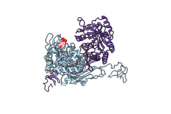
Cryo-Em Structure Of The Chsy3-Chpf1 Chondroitin Synthase Heterodimer
Organism: Human adenovirus sp., Homo sapiens
Method: ELECTRON MICROSCOPY Resolution:3.42 Å Release Date: 2026-03-04 Classification: SUGAR BINDING PROTEIN, TRANSFERASE Ligands: MN, UDP |
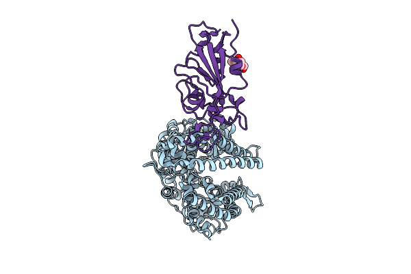 |
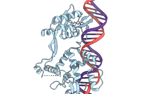
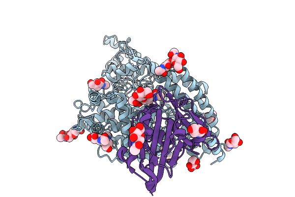
Crystal Structure Of Ratg13 Rbd Complexed With Sheep Ace2
Organism: Ovis aries, Bat coronavirus ratg13
Method: X-RAY DIFFRACTION Resolution:3.74 Å Release Date: 2025-11-26 Classification: VIRAL PROTEIN/HYDROLASE Ligands: NAG |
 |
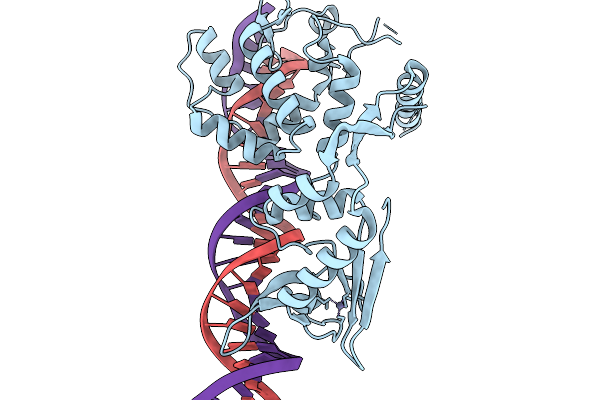
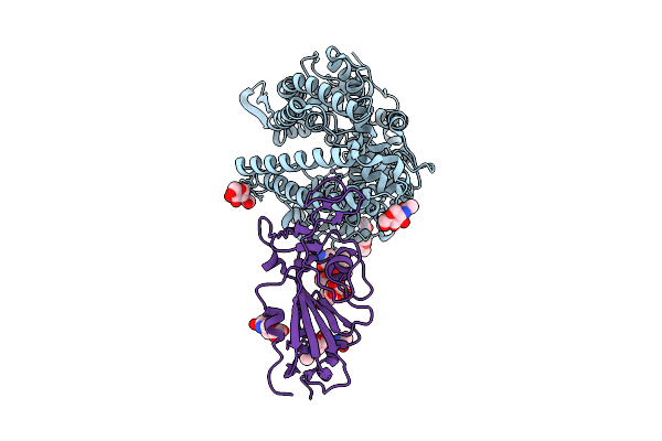
Crystal Structure Of Ratg13 Rbd Complexed With Cattle Ace2
Organism: Bos taurus, Bat coronavirus ratg13
Method: X-RAY DIFFRACTION Resolution:3.98 Å Release Date: 2025-11-26 Classification: VIRAL PROTEIN/HYDROLASE Ligands: NAG |
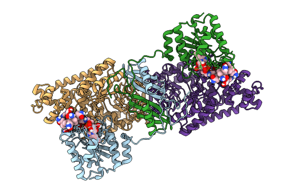 |
Open State Of Lysine 5,6-Aminomutase From Thermoanaerobacter Tengcongensis
Organism: Caldanaerobacter subterraneus subsp. tengcongensis
Method: ELECTRON MICROSCOPY Release Date: 2025-09-03 Classification: ISOMERASE Ligands: B12, 5AD |
 |
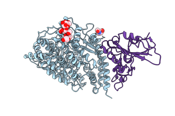
Eptesicus Fuscus Ace2 Peptidase Domain Bound To Vscov-A7 Rbd Complex
Organism: Eptesicus fuscus, Merbecovirus
Method: ELECTRON MICROSCOPY Release Date: 2025-08-27 Classification: HYDROLASE/VIRAL PROTEIN Ligands: NAG, ZN |
 |
Crystal Structure Of Ratg13 Receptor-Binding Domain Complexed With Squirrel Ace2
Organism: Petaurus norfolcensis, Bat coronavirus ratg13
Method: X-RAY DIFFRACTION Resolution:2.76 Å Release Date: 2025-08-06 Classification: VIRAL PROTEIN Ligands: CL, NAG, ZN |
 |
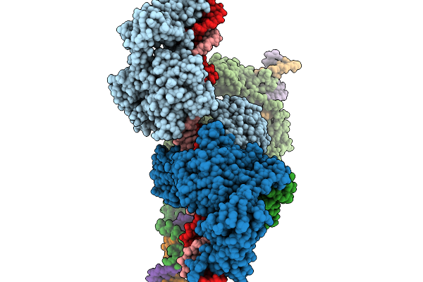
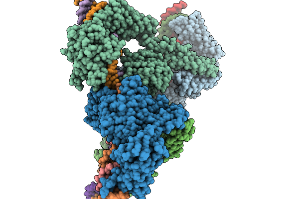
Structure Of The Sabia Virus Spike Complex In A Closed Conformation
Organism: Sabia virus
Method: ELECTRON MICROSCOPY Release Date: 2025-06-11 Classification: VIRAL PROTEIN Ligands: NAG, ZN |
 |
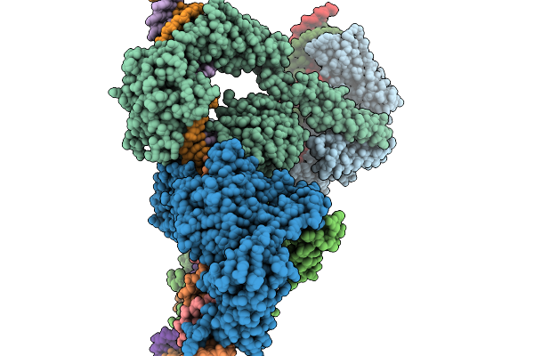
Structure Of The Sabia Virus Spike Complex In An Open Conformation
Organism: Sabia virus
Method: ELECTRON MICROSCOPY Release Date: 2025-06-11 Classification: VIRAL PROTEIN Ligands: NAG, K |
 |
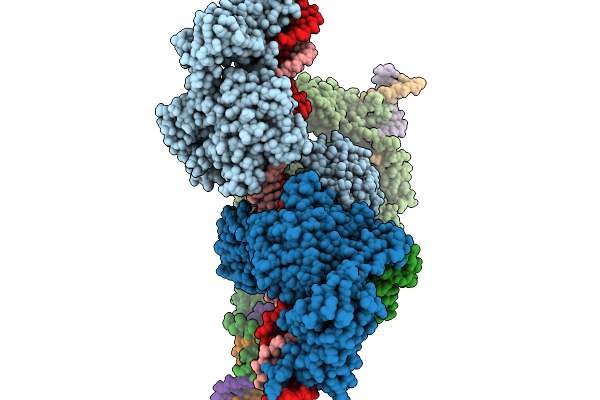
Structure Of The Sabia Virus Spike Complex H157M Mutant In A Closed Conformation
Organism: Sabia virus
Method: ELECTRON MICROSCOPY Release Date: 2025-06-11 Classification: VIRAL PROTEIN Ligands: NAG, ZN |
 |
Deletion Mutant Of Chitinase Mmchi60
Organism: Moritella marina
Method: X-RAY DIFFRACTION Resolution:2.69 Å Release Date: 2025-05-28 Classification: HYDROLASE |