
Search Count: 4,721
 |
Organism: Achromobacter xylosoxidans
Method: ELECTRON MICROSCOPY Release Date: 2026-06-03 Classification: MEMBRANE PROTEIN Ligands: HEM, CA |
 |
Crystal Structure Of A Flavin Dependent Baeyer Villiger Monooxygenase From Micromonospora Lupini Nbc_00409 In Complex With Fad
Organism: Micromonospora lupini
Method: X-RAY DIFFRACTION Resolution:3.20 Å Release Date: 2026-05-27 Classification: BIOSYNTHETIC PROTEIN Ligands: FAD |
 |
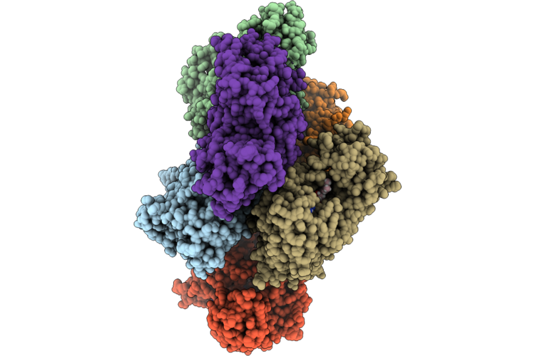
Cryoem Structure Of Native Quinol Dependent Nitric Oxide Reductase At Ph 8.0.
Organism: Achromobacter xylosoxidans
Method: ELECTRON MICROSCOPY Release Date: 2026-05-20 Classification: MEMBRANE PROTEIN Ligands: HEM, FE, CA, LMT, CU, UQ5 |
 |
Organism: Achromobacter xylosoxidans
Method: ELECTRON MICROSCOPY Release Date: 2026-05-20 Classification: MEMBRANE PROTEIN Ligands: HEM, FE, CA |
 |
Cryoem Structure Of Native Quinol Dependent Nitric Oxide Reductase With Hqn At Ph 6.5
Organism: Achromobacter xylosoxidans
Method: ELECTRON MICROSCOPY Resolution:2.30 Å Release Date: 2026-05-20 Classification: MEMBRANE PROTEIN Ligands: HEM, CA, FE, LMT, HQN, UQ5 |
 |
Cryoem Structure Of Native Quinol Dependent Nitric Oxide Reductase At Ph 8.0 On Gold Grid.
Organism: Achromobacter xylosoxidans
Method: ELECTRON MICROSCOPY Resolution:2.70 Å Release Date: 2026-05-06 Classification: MEMBRANE PROTEIN Ligands: HEM, CA, FE |
 |
Cryoem Structure Of Native Quinol Dependent Nitric Oxide Reductase Arg720Ala Variant At Ph 6.5 On Gold Grid.
Organism: Achromobacter xylosoxidans
Method: ELECTRON MICROSCOPY Resolution:2.90 Å Release Date: 2026-05-06 Classification: MEMBRANE PROTEIN Ligands: HEM, CA, FE |
 |
Cryoem Structure Of Native Quinol Dependent Nitric Oxide Reductase Trp718Ala Variant At Ph 6.5 On Gold Grid.
Organism: Achromobacter xylosoxidans
Method: ELECTRON MICROSCOPY Resolution:2.40 Å Release Date: 2026-05-06 Classification: MEMBRANE PROTEIN Ligands: HEM, CA, FE, LMT, UQ5 |
 |
Cryoem Structure Of Native Quinol Dependent Nitric Oxide Reductase Trp718Ala Variant With Quino At Ph 6.5 On Gold Grid.
Organism: Achromobacter xylosoxidans
Method: ELECTRON MICROSCOPY Resolution:2.40 Å Release Date: 2026-05-06 Classification: MEMBRANE PROTEIN Ligands: HEM, CA, FE, UQ5, HQE, LMT |
 |
Cryoem Structure Of Native Quinol Dependent Nitric Oxide Reductase At Ph 6.5
Organism: Achromobacter xylosoxidans
Method: ELECTRON MICROSCOPY Release Date: 2026-05-06 Classification: MEMBRANE PROTEIN Ligands: HEM, CA, FE |
 |
Cryoem Structure Of Native Quinol Dependent Nitric Oxide Reductase With Hqe At Ph 6.5
Organism: Achromobacter xylosoxidans
Method: ELECTRON MICROSCOPY Release Date: 2026-05-06 Classification: MEMBRANE PROTEIN Ligands: HEM, CA, FE, LMT, HQE, UQ5 |
 |
Organism: Amycolatopsis deserti
Method: X-RAY DIFFRACTION Resolution:2.08 Å Release Date: 2026-04-15 Classification: HYDROLASE |
 |
Crystal Structure Of The Polysaccharide Lyase Rbmb From Vibrio Cholerae Bound To Vibrio Polysaccharide
Organism: Vibrio cholerae
Method: X-RAY DIFFRACTION Resolution:1.62 Å Release Date: 2026-03-11 Classification: LYASE |
 |
Organism: Vibrio cholerae c6706
Method: X-RAY DIFFRACTION Resolution:2.10 Å Release Date: 2026-03-11 Classification: LYASE Ligands: GOL, CA |
 |
Cryoem Structure Of Aldehyde Dehydrogenase From Francisella Tularensis Subsp. Tularensis At 3.03A Resolution
Organism: Francisella tularensis subsp. tularensis
Method: ELECTRON MICROSCOPY Resolution:3.03 Å Release Date: 2026-01-28 Classification: OXIDOREDUCTASE |
 |
Cryo-Em Structure Of F-Atp Synthase From Mycobacteroides Abscessus (Rotational State 1)
Organism: Mycobacteroides abscessus subsp. abscessus
Method: ELECTRON MICROSCOPY Resolution:2.94 Å Release Date: 2026-01-14 Classification: HYDROLASE Ligands: ATP, MG, ADP |
 |
Cryo-Em Structure Of F-Atp Synthase From Mycobacteroides Abscessus (Rotational State 2)
Organism: Mycobacteroides abscessus subsp. abscessus
Method: ELECTRON MICROSCOPY Resolution:3.41 Å Release Date: 2026-01-14 Classification: HYDROLASE Ligands: ATP, MG, ADP |
 |
Cryo-Em Structure Of F-Atp Synthase From Mycobacteroides Abscessus (Rotational State 3)
Organism: Mycobacteroides abscessus subsp. abscessus
Method: ELECTRON MICROSCOPY Resolution:2.79 Å Release Date: 2026-01-14 Classification: HYDROLASE Ligands: ATP, MG, ADP |
 |
Cryo-Em Structure Of F-Atp Synthase C-Ring From Mycobacteroides Abscessus (Backbone)
Organism: Mycobacteroides abscessus subsp. abscessus
Method: ELECTRON MICROSCOPY Resolution:5.61 Å Release Date: 2026-01-14 Classification: HYDROLASE |
 |
Organism: Vibrio cholerae
Method: X-RAY DIFFRACTION Resolution:1.55 Å Release Date: 2025-07-02 Classification: SIGNALING PROTEIN Ligands: CL, ETA |