

Search Count: 19
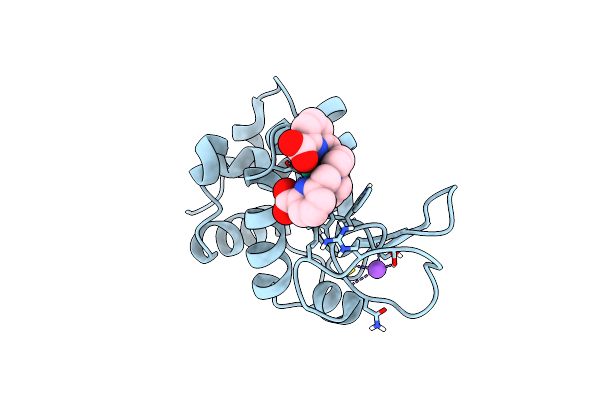 |
Crystal Structure Of Hen Egg White Lysozyme Co-Crystallized With 10 Mm Tbxo4
Organism: Gallus gallus
Method: X-RAY DIFFRACTION Resolution:1.70 Å Release Date: 2024-07-17 Classification: HYDROLASE Ligands: TB, 7MT, CL, NA |
Organism: Gallus gallus
Method: X-RAY DIFFRACTION
Release Date: 2024-07-17
Ligands: TB, 7MT, CL, NA
 |
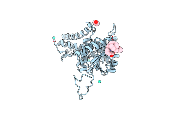
Crystal Structure Of N-Ethylmaleimide Reductase With Mutation Y187F (Nema Y187F) From Escherichia Coli
Organism: Escherichia coli
Method: X-RAY DIFFRACTION Resolution:2.30 Å Release Date: 2024-06-05 Classification: OXIDOREDUCTASE Ligands: FMN, 7MT |
Organism: Escherichia coli
Method: X-RAY DIFFRACTION
Release Date: 2024-06-05
Ligands: FMN, 7MT
 |
Pa14_16140 Protein: The Regulator Of An Operon Involved In The Biofilm Formation In Pa14 P. Aeruginosa
Organism: Pseudomonas aeruginosa ucbpp-pa14
Method: X-RAY DIFFRACTION Resolution:2.70 Å Release Date: 2024-02-28 Classification: CHAPERONE Ligands: GOL, 7MT |
Organism: Pseudomonas aeruginosa ucbpp-pa14
Method: X-RAY DIFFRACTION
Release Date: 2024-02-28
Ligands: GOL, 7MT
 |
Crystal Structure Of The Lactate Dehydrogenase Of Cyanobacterium Aponinum In Its Apo Form.
Organism: Cyanobacterium aponinum
Method: X-RAY DIFFRACTION Resolution:2.10 Å Release Date: 2022-08-03 Classification: OXIDOREDUCTASE Ligands: 7MT, TB, EDO |
Organism: Cyanobacterium aponinum
Method: X-RAY DIFFRACTION
Release Date: 2022-08-03
Ligands: 7MT, TB, EDO
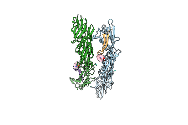 |
Crystal Structure Of Pita Fragment From Pilus Islet-2 Of Streptococcus Oralis With Tb-Xo4
Organism: Streptococcus oralis atcc 35037
Method: X-RAY DIFFRACTION Resolution:2.34 Å Release Date: 2022-07-13 Classification: CELL ADHESION Ligands: 7MT, TB |
Organism: Streptococcus oralis atcc 35037
Method: X-RAY DIFFRACTION
Release Date: 2022-07-13
Ligands: 7MT, TB
 |
Cryogenic Human Adiponectin Receptor 2 (Adipor2) With Tb-Xo4 Ligand Determined By Serial Crystallography (Ssx) Using Crystaldirect
Organism: Homo sapiens
Method: X-RAY DIFFRACTION Resolution:3.01 Å Release Date: 2021-05-12 Classification: MEMBRANE PROTEIN Ligands: ZN, OLA, OLB, 7MT |
Organism: Homo sapiens
Method: X-RAY DIFFRACTION
Release Date: 2021-05-12
Ligands: ZN, OLA, OLB, 7MT
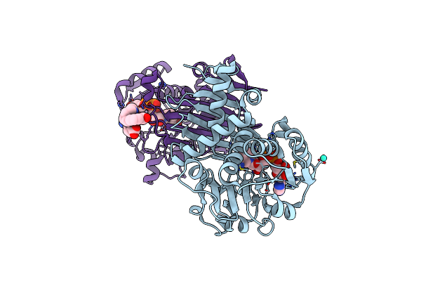 |
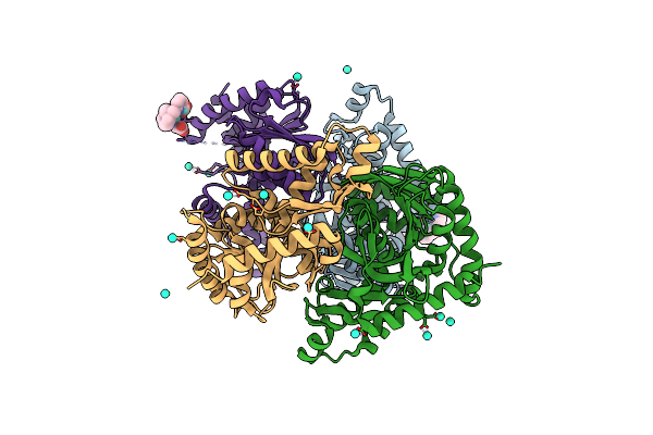
Crystal Structure Of The Iminosuccinate Reductase Of Paracoccus Denitrificans In Complex With Nad+
Organism: Paracoccus denitrificans (strain pd 1222)
Method: X-RAY DIFFRACTION Resolution:2.56 Å Release Date: 2019-08-14 Classification: OXIDOREDUCTASE Ligands: NAD, TB, 7MT |
Organism: Paracoccus denitrificans (strain pd 1222)
Method: X-RAY DIFFRACTION
Release Date: 2019-08-14
Ligands: NAD, TB, 7MT
 |
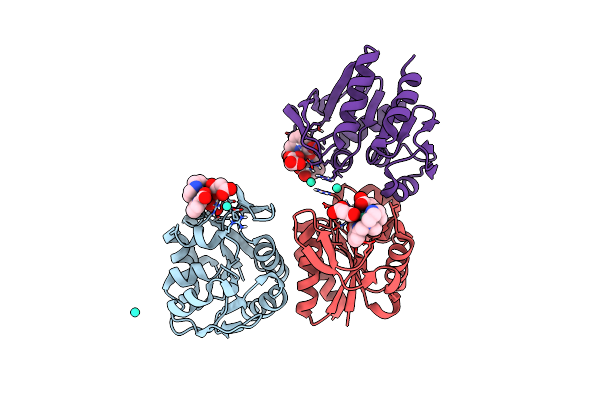
Crystal Structure Of Ignicoccus Islandicus Malate Dehydrogenase Co-Crystallized With 10 Mm Tb-Xo4
Organism: Ignicoccus islandicus dsm 13165
Method: X-RAY DIFFRACTION, SOLUTION SCATTERING Resolution:1.89 Å Release Date: 2019-07-17 Classification: OXIDOREDUCTASE Ligands: 7MT, TB, CL |
Organism: Ignicoccus islandicus dsm 13165
Method: X-RAY DIFFRACTION, SOLUTION SCATTERING
Release Date: 2019-07-17
Ligands: 7MT, TB, CL
 |
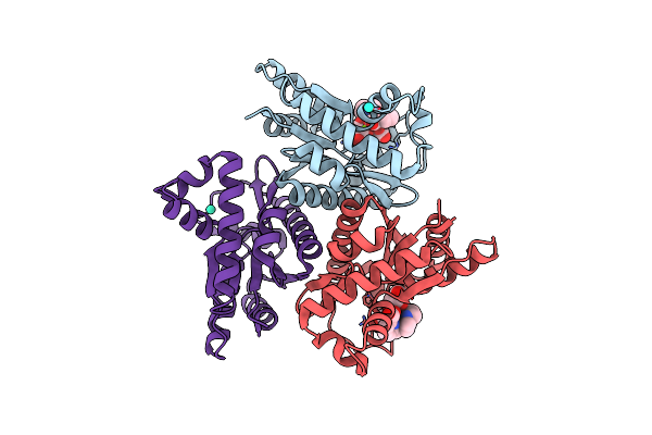
Crystal Structure Of The Protease 1 (E29A,E60A,E80A) From Pyrococcus Horikoshii Co-Crystallized With Tb-Xo4.
Organism: Pyrococcus horikoshii (strain atcc 700860 / dsm 12428 / jcm 9974 / nbrc 100139 / ot-3)
Method: X-RAY DIFFRACTION Resolution:2.00 Å Release Date: 2019-06-19 Classification: HYDROLASE Ligands: 7MT, MLI, TB |
Organism: Pyrococcus horikoshii (strain atcc 700860 / dsm 12428 / jcm 9974 / nbrc 100139 / ot-3)
Method: X-RAY DIFFRACTION
Release Date: 2019-06-19
Ligands: 7MT, MLI, TB
 |
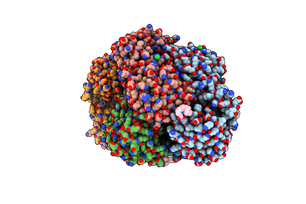
Crystal Structure Of The Adenylate Kinase From Methanothermococcus Thermolithotrophicus Co-Crystallized With Tb-Xo4
Organism: Methanothermococcus thermolithotrophicus
Method: X-RAY DIFFRACTION Resolution:1.96 Å Release Date: 2019-06-19 Classification: TRANSFERASE Ligands: 7MT, TB, MG, GOL |
Organism: Methanothermococcus thermolithotrophicus
Method: X-RAY DIFFRACTION
Release Date: 2019-06-19
Ligands: 7MT, TB, MG, GOL
 |
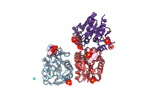
Crystal Structure Of Coenzyme F420H2 Oxidase (Fpra) Co-Crystallized With 10 Mm Tb-Xo4
Organism: Methanothermococcus thermolithotrophicus
Method: X-RAY DIFFRACTION Resolution:2.20 Å Release Date: 2018-10-31 Classification: OXIDOREDUCTASE Ligands: FMN, 7MT, TB, CL, FE |
Organism: Methanothermococcus thermolithotrophicus
Method: X-RAY DIFFRACTION
Release Date: 2018-10-31
Ligands: FMN, 7MT, TB, CL, FE
 |
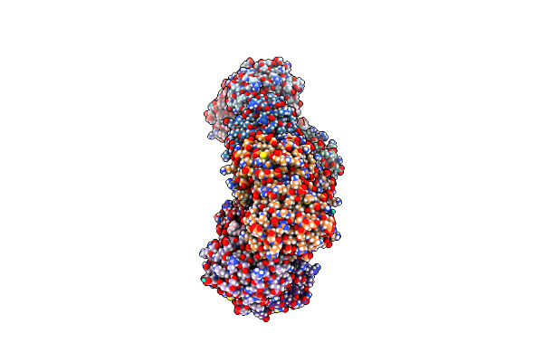
Crystal Structure Of Protease 1 From Pyrococcus Horikoshii Co-Cystallized In Presence Of 10 Mm Tb-Xo4 And Ammonium Sulfate.
Organism: Pyrococcus horikoshii
Method: X-RAY DIFFRACTION Resolution:1.65 Å Release Date: 2018-10-03 Classification: CELL CYCLE Ligands: 7MT, SO4, TB, 2HA |
Organism: Pyrococcus horikoshii
Method: X-RAY DIFFRACTION
Release Date: 2018-10-03
Ligands: 7MT, SO4, TB, 2HA
 |
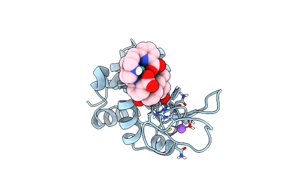
Structure Of Protease 1 From Pyrococcus Horikoshii Co-Crystallized In Presence Of 10 Mm Tb-Xo4 And Potassium Iodide.
Organism: Pyrococcus horikoshii
Method: X-RAY DIFFRACTION Resolution:2.19 Å Release Date: 2018-10-03 Classification: HYDROLASE Ligands: 7MT, IOD, TB |
Organism: Pyrococcus horikoshii
Method: X-RAY DIFFRACTION
Release Date: 2018-10-03
Ligands: 7MT, IOD, TB
 |
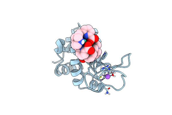
Crystal Structure Of Hen Egg-White Lysozyme Co-Crystallized In Presence Of 100 Mm Tb-Xo4
Organism: Gallus gallus
Method: X-RAY DIFFRACTION Resolution:1.20 Å Release Date: 2018-10-03 Classification: HYDROLASE Ligands: TB, 7MT, CL, NA, ACT |
Organism: Gallus gallus
Method: X-RAY DIFFRACTION
Release Date: 2018-10-03
Ligands: TB, 7MT, CL, NA, ACT
 |
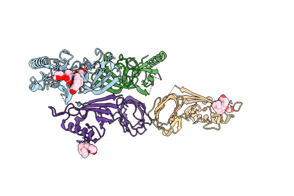
Crystal Structure Of Hen Egg-White Lysozyme Co-Crystallized In Presence Of 100 Mm Tb-Xo4 And 100 Mm Sodium Sulfate
Organism: Gallus gallus
Method: X-RAY DIFFRACTION Resolution:1.30 Å Release Date: 2018-10-03 Classification: HYDROLASE Ligands: TB, CL, NA, 7MT |
Organism: Gallus gallus
Method: X-RAY DIFFRACTION
Release Date: 2018-10-03
Ligands: TB, CL, NA, 7MT
 |
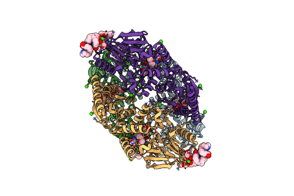
Structure Of The Bacteriophage T5 Distal Tail Protein Pb9 Co-Crystallized With 10Mm Tb-Xo4
Organism: Escherichia phage t5
Method: X-RAY DIFFRACTION Resolution:1.80 Å Release Date: 2018-10-03 Classification: VIRAL PROTEIN Ligands: 7MT, EDO, CL, NA, TB |
Organism: Escherichia phage t5
Method: X-RAY DIFFRACTION
Release Date: 2018-10-03
Ligands: 7MT, EDO, CL, NA, TB
 |
Structure Of F420H2 Oxidase (Fpra) Co-Crystallized With 10Mm Tb-Xo4 And Calcium Chloride
Organism: Methanothermococcus thermolithotrophicus
Method: X-RAY DIFFRACTION Resolution:1.74 Å Release Date: 2018-10-03 Classification: OXIDOREDUCTASE Ligands: FMN, CA, TB, GOL, PE4, 7MT, NA |
Organism: Methanothermococcus thermolithotrophicus
Method: X-RAY DIFFRACTION
Release Date: 2018-10-03
Ligands: FMN, CA, TB, GOL, PE4, 7MT, NA
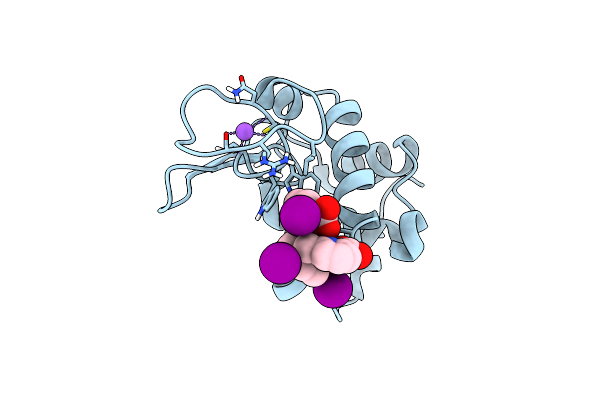 |
Crystal Structure Of Hen Egg-White Lysozyme Co-Crystallized In Presence Of 100 Mm Tb-Xo4 And 100 Mm Potassium Iodide.
Organism: Gallus gallus
Method: X-RAY DIFFRACTION Resolution:1.42 Å Release Date: 2018-10-03 Classification: HYDROLASE Ligands: TB, IOD, NA, 7MT |
Organism: Gallus gallus
Method: X-RAY DIFFRACTION
Release Date: 2018-10-03
Ligands: TB, IOD, NA, 7MT
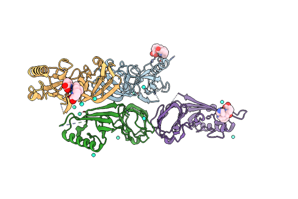 |
Bacteriophage T5 Distal Tail Protein Pb9 Co-Crystallized With Tb-Xo4
Organism: Escherichia phage t5
Method: X-RAY DIFFRACTION Resolution:2.00 Å Release Date: 2017-09-20 Classification: VIRAL PROTEIN Ligands: TB, 7MT, CL |
Organism: Escherichia phage t5
Method: X-RAY DIFFRACTION
Release Date: 2017-09-20
Ligands: TB, 7MT, CL