
Search Count: 85
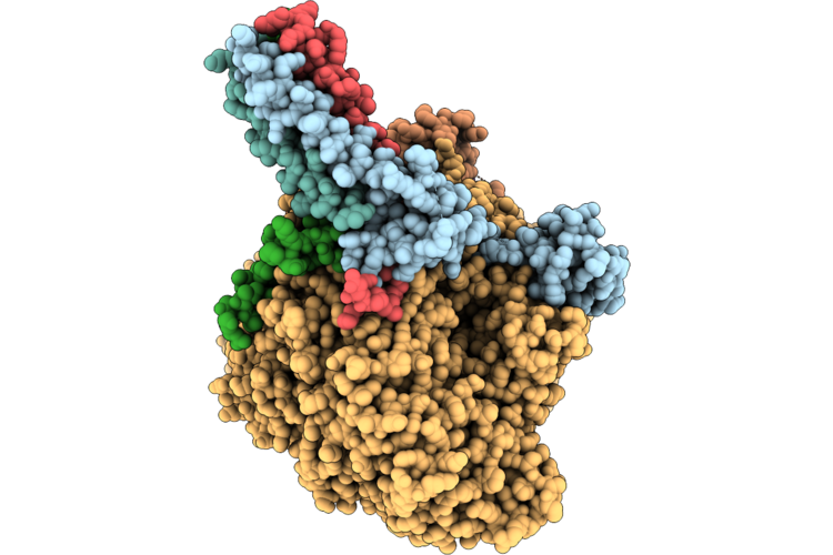 |
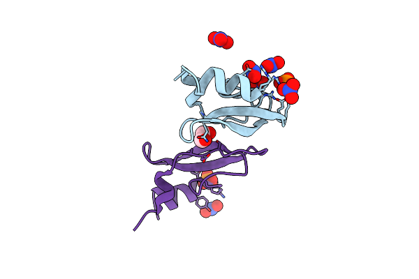
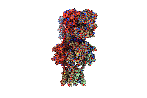
Cryo-Em Structure Of The Human Measles Virus Rna-Dependent Rna Polymerase Complex Bound To Viral Protein C
Organism: Escherichia coli k-12, Measles virus genotype a-vaccine, Synthetic construct, Aequorea victoria
Method: ELECTRON MICROSCOPY Release Date: 2026-05-27 Classification: VIRAL PROTEIN Ligands: ZN |
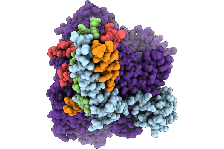 |
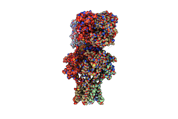
Organism: Escherichia coli k-12, Measles virus genotype a-vaccine, Synthetic construct
Method: ELECTRON MICROSCOPY Resolution:2.60 Å Release Date: 2026-05-27 Classification: VIRAL PROTEIN Ligands: ZN |
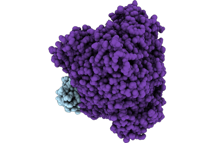 |
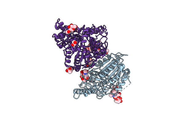
Cryo-Em Structure Of The Human Measles Virus Rna-Dependent Rna Polymerase Complex
Organism: Escherichia coli k-12, Measles virus genotype a-vaccine, Synthetic construct
Method: ELECTRON MICROSCOPY Resolution:3.17 Å Release Date: 2026-05-27 Classification: VIRAL PROTEIN Ligands: ZN |
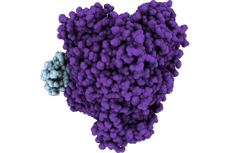 |
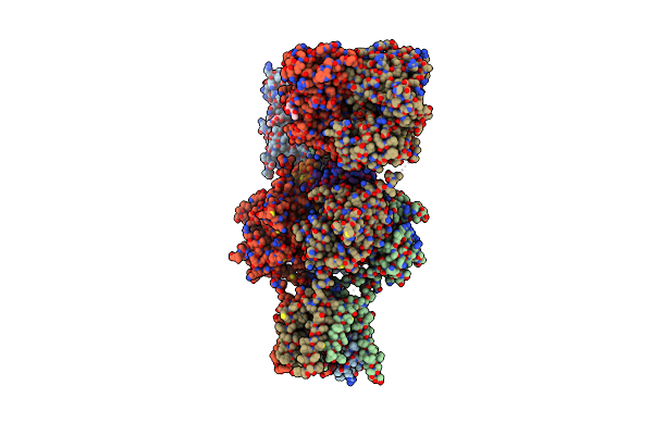
Cryo-Em Structure Of The Human Measles Virus Rna-Dependent Rna Polymerase Bound To Allosteric Inhibitor Erdrp-0519
Organism: Escherichia coli k-12, Measles virus genotype a-vaccine, Synthetic construct
Method: ELECTRON MICROSCOPY Resolution:3.13 Å Release Date: 2026-05-27 Classification: VIRAL PROTEIN Ligands: ZN, A1EF9 |
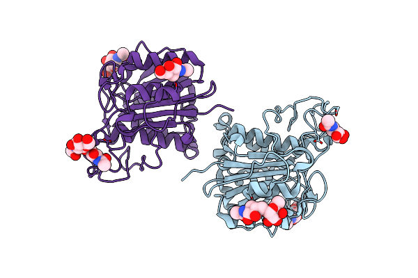 |
The Crystal Structure Of Vypal2-C214A, A Dead Mutant Of Vypal2 Peptide Asparaginyl Ligase In Form Ii
Organism: Viola philippica
Method: X-RAY DIFFRACTION Resolution:1.80 Å Release Date: 2022-07-13 Classification: PLANT PROTEIN Ligands: NAG |
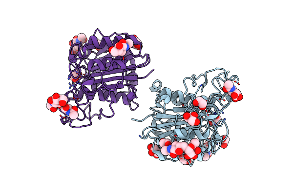 |
The Crystal Structure Of Vypal2-I244V, A More Efficient Mutant Of Vypal2 Peptide Asparaginyl Ligase In Its Active Enzyme Form
Organism: Viola philippica
Method: X-RAY DIFFRACTION Resolution:1.59 Å Release Date: 2022-06-29 Classification: PLANT PROTEIN Ligands: NAG, EDO, GOL |
 |
The Crystal Structure Of Vypal2-C214A, A Dead Mutant Of Vypal2 Peptide Asparaginyl Ligase In Form I
Organism: Viola philippica
Method: X-RAY DIFFRACTION Resolution:1.90 Å Release Date: 2022-06-29 Classification: PLANT PROTEIN |
 |
The Crystal Structure Of Vypal2 Peptide Asparaginyl Ligase In Its Active Enzyme Form
Organism: Viola philippica
Method: X-RAY DIFFRACTION Resolution:2.30 Å Release Date: 2022-06-29 Classification: PLANT PROTEIN Ligands: NAG, EDO |
 |
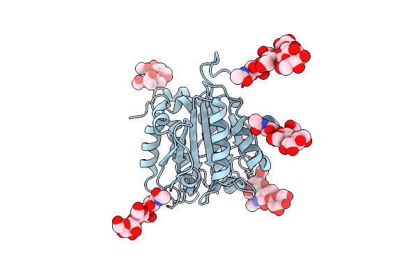
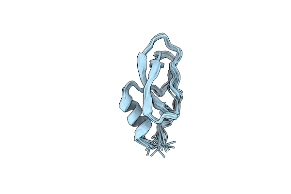
Pore-Modulating Toxins Exploit Inherent Slow Inactivation To Block K+ Channels
Organism: Conus striatus
Method: X-RAY DIFFRACTION Resolution:1.30 Å Release Date: 2019-08-21 Classification: TOXIN Ligands: SO4 |
 |
Pore-Modulating Toxins Exploit Inherent Slow Inactivation To Block K+ Channels
Organism: Conus striatus
Method: X-RAY DIFFRACTION Resolution:1.30 Å Release Date: 2019-08-21 Classification: TOXIN Ligands: PO4, NO3, EDO |
 |
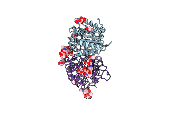
Organism: Viola philippica
Method: X-RAY DIFFRACTION Resolution:2.40 Å Release Date: 2019-05-15 Classification: LIGASE Ligands: NAG, EDO, PEG |
 |
Crystal Structure Of Glua2 A622T, Con-Ikot-Ikot Snail Toxin, Partial Agonist Ka And Postitive Modulator (R,R)-2B Complex
Organism: Rattus norvegicus, Conus striatus
Method: X-RAY DIFFRACTION Resolution:3.50 Å Release Date: 2014-08-13 Classification: Transport protein/toxin Ligands: KAI, NAG, FWF |
 |
Crystal Structure Of Glua2, Con-Ikot-Ikot Snail Toxin, Partial Agonist Fw And Postitive Modulator (R,R)-2B Complex
Organism: Rattus norvegicus, Conus striatus
Method: X-RAY DIFFRACTION Resolution:3.69 Å Release Date: 2014-08-13 Classification: Transport protein/toxin Ligands: NAG, FWD, FWF |
 |
Crystal Structure Of Glua2, Con-Ikot-Ikot Snail Toxin, Partial Agonist Ka And Postitive Modulator (R,R)-2B Complex
Organism: Rattus norvegicus, Conus striatus
Method: X-RAY DIFFRACTION Resolution:3.58 Å Release Date: 2014-08-13 Classification: Transport protein/toxin Ligands: KAI, NAG, FWF |
 |
Crystal Structure Of Glua2 T625G, Con-Ikot-Ikot Snail Toxin, Partial Agonist Ka And Postitive Modulator (R,R)-2B Complex
Organism: Rattus norvegicus, Conus striatus
Method: X-RAY DIFFRACTION Resolution:3.51 Å Release Date: 2014-08-13 Classification: Transport protein/Toxin Ligands: KAI, NAG, FWF |
 |
Crystal Structure Of Glua2, Con-Ikot-Ikot Snail Toxin, Partial Agonist Ka And Postitive Modulator (R,R)-2B Complex, Glua2Cryst2 Construct
Organism: Rattus norvegicus, Conus striatus
Method: X-RAY DIFFRACTION Resolution:3.70 Å Release Date: 2014-08-13 Classification: Transport protein/Toxin Ligands: KAI, NAG, FWF |
 |
Organism: Conus striatus
Method: X-RAY DIFFRACTION Resolution:2.20 Å Release Date: 2014-08-13 Classification: TOXIN Ligands: ZN |
 |
Organism: Conus striatus
Method: X-RAY DIFFRACTION Resolution:1.58 Å Release Date: 2014-08-13 Classification: TOXIN |
 |
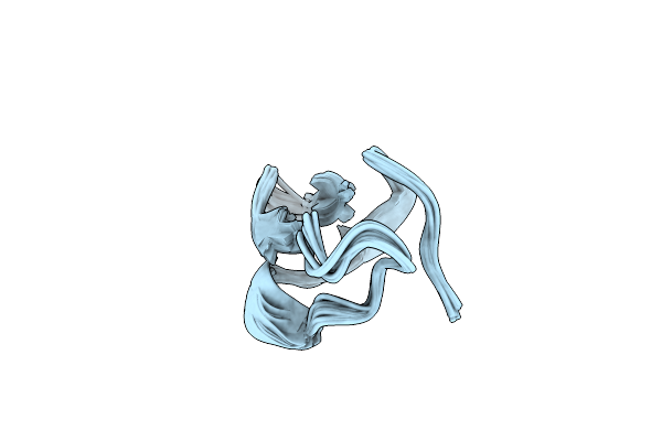
Conkunitzin-S1 Is The First Member Of A New Kunitz-Type Neurotoxin Family- Structural And Functional Characterization
|
 |