
Search Count: 3,105
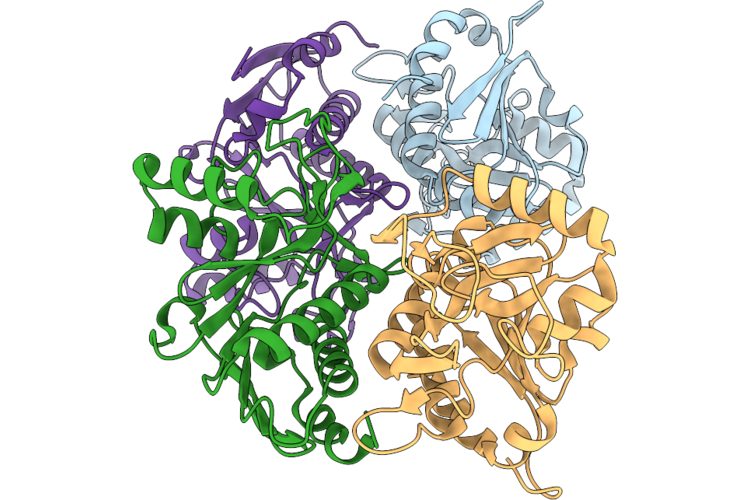 |
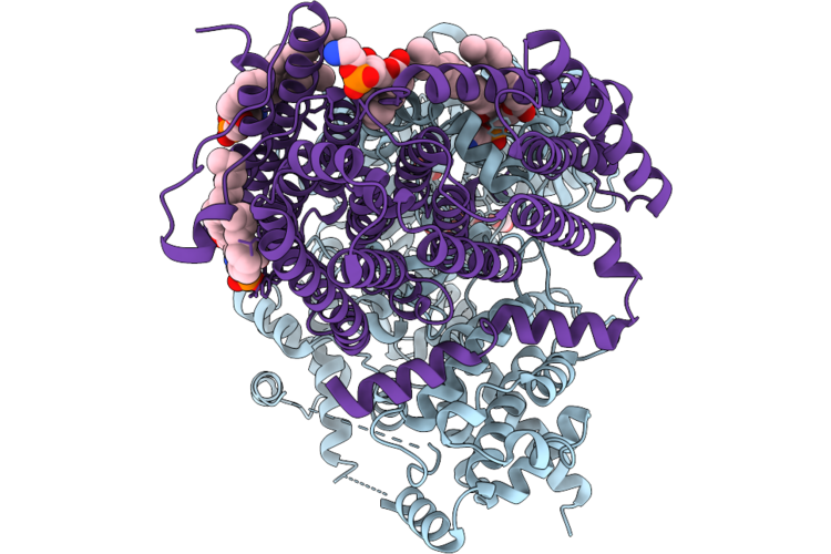
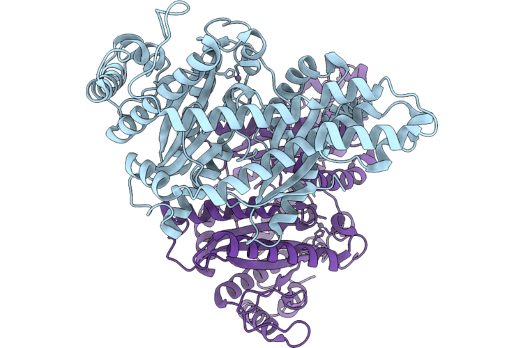
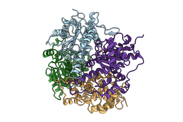
Crystal Structure Of Thoeris Protein Thsa
Organism: Bacillus velezensis yau b9601-y2
Method: X-RAY DIFFRACTION Resolution:2.59 Å Release Date: 2026-05-13 Classification: HYDROLASE |
Organism: Bacillus velezensis yau b9601-y2
Method: X-RAY DIFFRACTION
Release Date: 2026-05-13
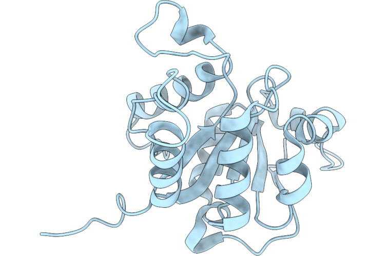 |
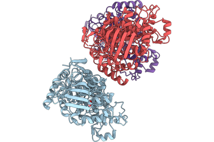
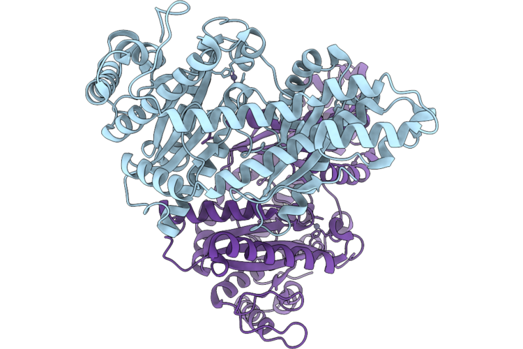
Crystal Structure Of Thoeris Protein Thsb
Organism: Bacillus velezensis yau b9601-y2
Method: X-RAY DIFFRACTION Resolution:1.58 Å Release Date: 2026-05-13 Classification: HYDROLASE |
Organism: Bacillus velezensis yau b9601-y2
Method: X-RAY DIFFRACTION
Release Date: 2026-05-13
 |
Structure Of Daba2B2 Complex Solved Under Ambient Condition
Organism: Halothiobacillus neapolitanus c2
Method: ELECTRON MICROSCOPY Release Date: 2026-04-29 Classification: TRANSPORT PROTEIN Ligands: ZN, CO2, 6OU |
Organism: Halothiobacillus neapolitanus c2
Method: ELECTRON MICROSCOPY
Release Date: 2026-04-29
Ligands: ZN, CO2, 6OU
 |
Structure Of Daba2B2 Complex Under 0.1M Bicarbonate
Organism: Halothiobacillus neapolitanus c2
Method: ELECTRON MICROSCOPY Release Date: 2026-04-29 Classification: TRANSPORT PROTEIN Ligands: ZN, BCT, CO2, 6OU |
Organism: Halothiobacillus neapolitanus c2
Method: ELECTRON MICROSCOPY
Release Date: 2026-04-29
Ligands: ZN, BCT, CO2, 6OU
 |
Structure Of Daba2B2 Complex Under 17 Mm Co2
Organism: Halothiobacillus neapolitanus c2
Method: ELECTRON MICROSCOPY Release Date: 2026-04-29 Classification: TRANSPORT PROTEIN Ligands: ZN, CO2, 6OU |
Organism: Halothiobacillus neapolitanus c2
Method: ELECTRON MICROSCOPY
Release Date: 2026-04-29
Ligands: ZN, CO2, 6OU
 |
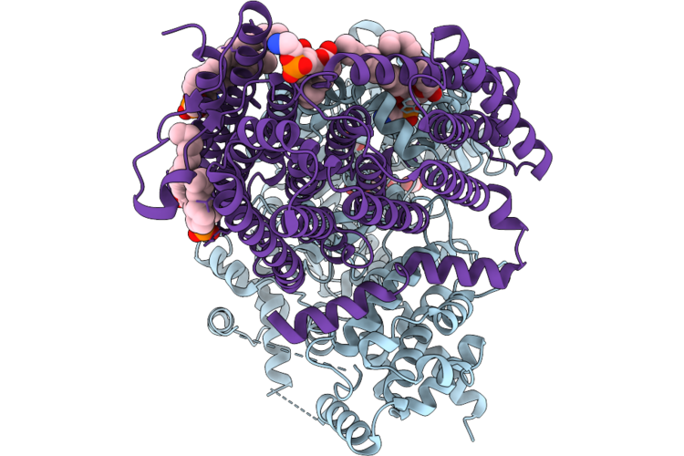
Crystal Structure Of Apo Abghmp
Organism: Acidobacteriota
Method: X-RAY DIFFRACTION Resolution:2.16 Å Release Date: 2026-04-29 Classification: TRANSFERASE Ligands: ACT |
Organism: Acidobacteriota
Method: X-RAY DIFFRACTION
Release Date: 2026-04-29
Ligands: ACT
 |
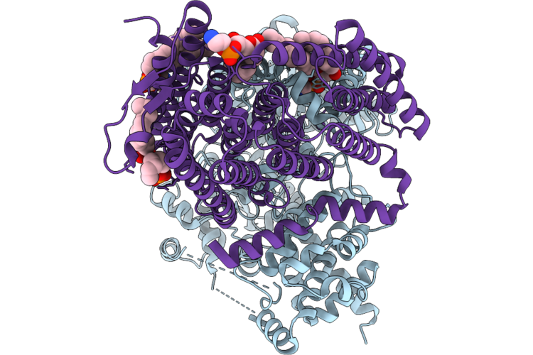
Crystal Structure Of Abghmp In Complex With L-Ara And Amppnp
Organism: Acidobacteriota
Method: X-RAY DIFFRACTION Resolution:2.60 Å Release Date: 2026-04-29 Classification: TRANSFERASE Ligands: ANP, ARB, MG |
Organism: Acidobacteriota
Method: X-RAY DIFFRACTION
Release Date: 2026-04-29
Ligands: ANP, ARB, MG
 |
The Cryo-Em Structure Of Zea Mays Gln1
Organism: Zea mays
Method: ELECTRON MICROSCOPY Release Date: 2026-04-29 Classification: PLANT PROTEIN |
Organism: Zea mays
Method: ELECTRON MICROSCOPY
Release Date: 2026-04-29
 |
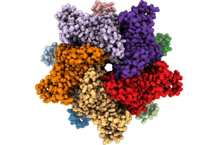
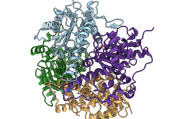
Cryo-Em Structure Of H. Neapolitanus Csosca In Oxidizing Conditions, Hexamer
Organism: Escherichia coli k-12, Halothiobacillus neapolitanus c2
Method: ELECTRON MICROSCOPY Resolution:2.13 Å Release Date: 2026-04-22 Classification: LYASE Ligands: ZN |
Organism: Escherichia coli k-12, Halothiobacillus neapolitanus c2
Method: ELECTRON MICROSCOPY
Release Date: 2026-04-22
Ligands: ZN
 |
Cryo-Em Structure Of H. Neapolitanus Csosca In Oxidizing Conditions, Dimer, Major State, Active Conformation
Organism: Escherichia coli k-12, Halothiobacillus neapolitanus c2
Method: ELECTRON MICROSCOPY Resolution:2.15 Å Release Date: 2026-04-22 Classification: LYASE Ligands: ZN |
Organism: Escherichia coli k-12, Halothiobacillus neapolitanus c2
Method: ELECTRON MICROSCOPY
Release Date: 2026-04-22
Ligands: ZN
 |
Cryo-Em Structure Of H. Neapolitanus Csosca In Oxidizing Conditions, Dimer, Minor State
Organism: Escherichia coli k-12, Halothiobacillus neapolitanus c2
Method: ELECTRON MICROSCOPY Resolution:2.45 Å Release Date: 2026-04-22 Classification: LYASE Ligands: ZN |
Organism: Escherichia coli k-12, Halothiobacillus neapolitanus c2
Method: ELECTRON MICROSCOPY
Release Date: 2026-04-22
Ligands: ZN
 |
Cryo-Em Structure Of H. Neapolitanus Csosca In Reducing Conditions, Hexamer
Organism: Escherichia coli k-12, Halothiobacillus neapolitanus c2
Method: ELECTRON MICROSCOPY Resolution:2.06 Å Release Date: 2026-04-22 Classification: LYASE Ligands: ZN |
Organism: Escherichia coli k-12, Halothiobacillus neapolitanus c2
Method: ELECTRON MICROSCOPY
Release Date: 2026-04-22
Ligands: ZN
 |
Cryo-Em Structure Of H. Neapolitanus Csosca In Reducing Conditions, Dimer, Major State, Inactive Conformation
Organism: Escherichia coli k-12, Halothiobacillus neapolitanus c2
Method: ELECTRON MICROSCOPY Resolution:2.12 Å Release Date: 2026-04-22 Classification: LYASE Ligands: ZN |
Organism: Escherichia coli k-12, Halothiobacillus neapolitanus c2
Method: ELECTRON MICROSCOPY
Release Date: 2026-04-22
Ligands: ZN
 |
Cryo-Em Structure Of H. Neapolitanus Csosca In Reducing Conditions, Dimer, Minor State
Organism: Escherichia coli k-12, Halothiobacillus neapolitanus c2
Method: ELECTRON MICROSCOPY Resolution:2.27 Å Release Date: 2026-04-22 Classification: LYASE Ligands: ZN |
Organism: Escherichia coli k-12, Halothiobacillus neapolitanus c2
Method: ELECTRON MICROSCOPY
Release Date: 2026-04-22
Ligands: ZN
 |
Cryo-Em Structure Of H. Neapolitanus Csosca C283A/C284A Inactive Mutant, Hexamer
Organism: Escherichia coli k-12, Halothiobacillus neapolitanus c2
Method: ELECTRON MICROSCOPY Resolution:2.08 Å Release Date: 2026-04-22 Classification: LYASE Ligands: ZN |
Organism: Escherichia coli k-12, Halothiobacillus neapolitanus c2
Method: ELECTRON MICROSCOPY
Release Date: 2026-04-22
Ligands: ZN
 |
Cryo-Em Structure Of H. Neapolitanus Csosca C283A/C284A Inactive Mutant, Dimer, State 1
Organism: Escherichia coli k-12, Halothiobacillus neapolitanus c2
Method: ELECTRON MICROSCOPY Resolution:2.18 Å Release Date: 2026-04-22 Classification: LYASE Ligands: ZN |
Organism: Escherichia coli k-12, Halothiobacillus neapolitanus c2
Method: ELECTRON MICROSCOPY
Release Date: 2026-04-22
Ligands: ZN
 |
Cryo-Em Structure Of H. Neapolitanus Csosca C283A/C284A Inactive Mutant, Dimer, State 2
Organism: Escherichia coli k-12, Halothiobacillus neapolitanus c2
Method: ELECTRON MICROSCOPY Resolution:2.22 Å Release Date: 2026-04-22 Classification: LYASE Ligands: ZN |
Organism: Escherichia coli k-12, Halothiobacillus neapolitanus c2
Method: ELECTRON MICROSCOPY
Release Date: 2026-04-22
Ligands: ZN
 |
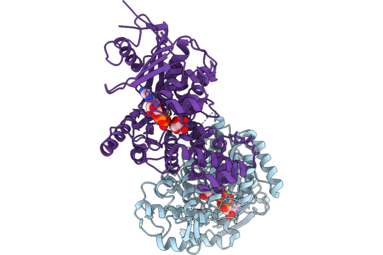
Three Viral Endonucleases Bound To The Same Inhibitor (9-Hydroxy-3,4-Dihydro-2H-Pyrazino[1,2-C]Pyrimidine-1,8-Dione Derivative). (2) La Crosse Virus L Protein Endonuclease With Manganese Ions.
Organism: La crosse virus
Method: X-RAY DIFFRACTION Resolution:2.80 Å Release Date: 2026-04-15 Classification: RNA BINDING PROTEIN Ligands: MN, A1J4F, SO4 |
Organism: La crosse virus
Method: X-RAY DIFFRACTION
Release Date: 2026-04-15
Ligands: MN, A1J4F, SO4
 |
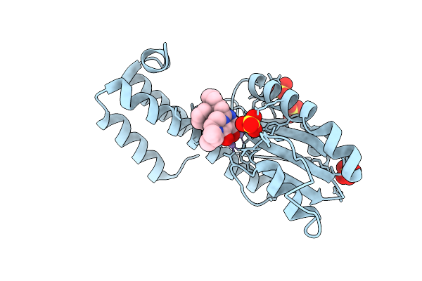
Crystal Structure Of None-Heme Iron Enzyme (Tqam) From Trichoderma Atroviride Bound With Iron And 2-Aminoisobutyric Acid
Organism: Trichoderma atroviride
Method: X-RAY DIFFRACTION Resolution:2.15 Å Release Date: 2026-02-04 Classification: METAL BINDING PROTEIN Ligands: FE, AIB |
Organism: Trichoderma atroviride
Method: X-RAY DIFFRACTION
Release Date: 2026-02-04
Ligands: FE, AIB
 |
Crystal Structure Of None-Heme Iron Enzyme (Tqam) From Trichoderma Atroviride Bound With Iron
Organism: Trichoderma atroviride
Method: X-RAY DIFFRACTION Resolution:2.60 Å Release Date: 2026-01-28 Classification: METAL BINDING PROTEIN Ligands: FE |
Organism: Trichoderma atroviride
Method: X-RAY DIFFRACTION
Release Date: 2026-01-28
Ligands: FE