

Search Count: 184
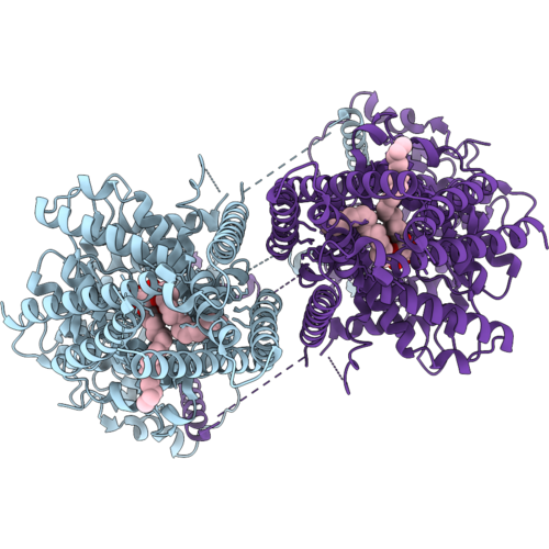 |
Structure Of Class Ii Chitin Synthase, Chs1, From Candida Albicans (Cachs1) In The Apo State
Organism: Candida albicans
Method: ELECTRON MICROSCOPY Resolution:3.02 Å Release Date: 2026-10-14 Classification: MEMBRANE PROTEIN Ligands: PEE |
Organism: Candida albicans
Method: ELECTRON MICROSCOPY
Release Date: 2026-10-14
Ligands: PEE
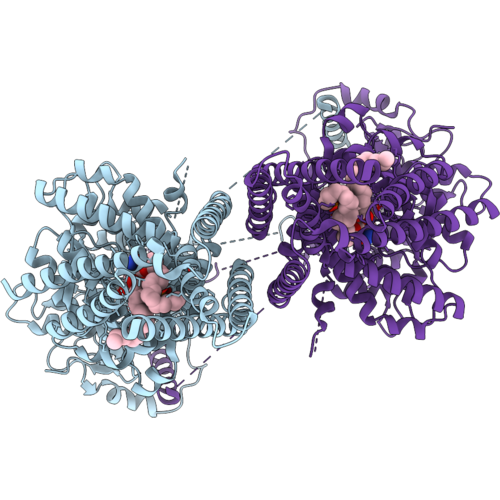 |
Structure Of Class Ii Chitin Synthase, Chs1, From Candida Albicans (Cachs1), State 1 (Post Udp Hydrolysis) After 5 Min Reaction
Organism: Candida albicans
Method: ELECTRON MICROSCOPY Resolution:3.38 Å Release Date: 2026-10-14 Classification: MEMBRANE PROTEIN Ligands: MG, PEE, NAG, UDP |
Organism: Candida albicans
Method: ELECTRON MICROSCOPY
Release Date: 2026-10-14
Ligands: MG, PEE, NAG, UDP
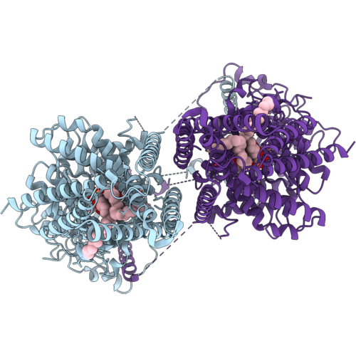 |
Structure Of Class Ii Chitin Synthase, Chs1, From Candida Albicans (Cachs1), State 2 (Pre-Translocation) After 5 Min Reaction
Organism: Candida albicans
Method: ELECTRON MICROSCOPY Release Date: 2026-10-14 Classification: MEMBRANE PROTEIN Ligands: UDP, PEE, MG |
Organism: Candida albicans
Method: ELECTRON MICROSCOPY
Release Date: 2026-10-14
Ligands: UDP, PEE, MG
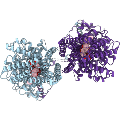 |
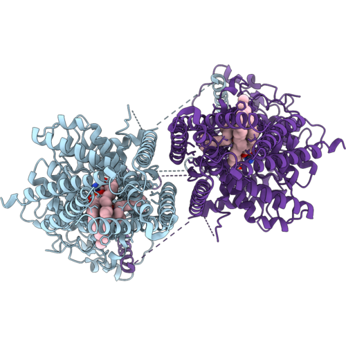
Structure Of Class Ii Chitin Synthase, Chs1, From Candida Albicans (Cachs1), State 3 (Post-Translocation) After 5 Min Reaction
Organism: Candida albicans
Method: ELECTRON MICROSCOPY Release Date: 2026-10-14 Classification: MEMBRANE PROTEIN Ligands: UDP, PEE |
Organism: Candida albicans
Method: ELECTRON MICROSCOPY
Release Date: 2026-10-14
Ligands: UDP, PEE
 |
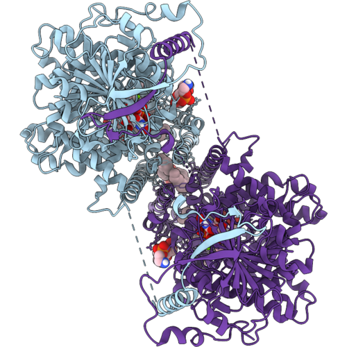
Structure Of Class Ii Chitin Synthase, Chs1, From Candida Albicans (Cachs1), Bound To Diynyl Arylamine (Da, Aka Ro-09-3143)
Organism: Candida albicans
Method: ELECTRON MICROSCOPY Release Date: 2026-10-14 Classification: MEMBRANE PROTEIN Ligands: PEE, A1DEH |
Organism: Candida albicans
Method: ELECTRON MICROSCOPY
Release Date: 2026-10-14
Ligands: PEE, A1DEH
 |
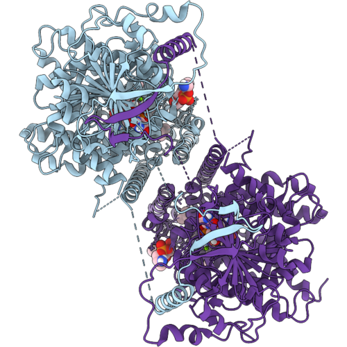
Candida Albicans Chitin Synthase 1 (Cachs1) Bound To Udp-Glcnac And Diynyl Arylamine (Da, Aka Ro-09-3143)
Organism: Candida albicans
Method: ELECTRON MICROSCOPY Resolution:2.96 Å Release Date: 2026-10-14 Classification: MEMBRANE PROTEIN Ligands: PEE, MG, A1DEH, UD1 |
Organism: Candida albicans
Method: ELECTRON MICROSCOPY
Release Date: 2026-10-14
Ligands: PEE, MG, A1DEH, UD1
 |
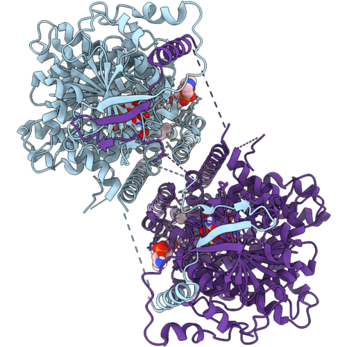
Candida Albicans Chitin Synthase 1 (Cachs1) Intermediate State (Da-2), Bound To Udp And Diynyl Arylamine (Da, Aka Ro-09-3143)
Organism: Candida albicans
Method: ELECTRON MICROSCOPY Resolution:3.02 Å Release Date: 2026-10-14 Classification: MEMBRANE PROTEIN Ligands: PEE, MG, A1DEH, UDP |
Organism: Candida albicans
Method: ELECTRON MICROSCOPY
Release Date: 2026-10-14
Ligands: PEE, MG, A1DEH, UDP
 |
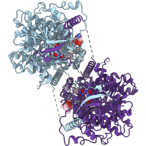
Candida Albicans Chitin Synthase 1 (Cachs1) Intermediate State (Da-3), Bound To Udp And Diynyl Arylamine (Da, Aka Ro-09-3143)
Organism: Candida albicans
Method: ELECTRON MICROSCOPY Resolution:3.00 Å Release Date: 2026-10-14 Classification: MEMBRANE PROTEIN Ligands: PEE, MG, A1DEH, UDP |
Organism: Candida albicans
Method: ELECTRON MICROSCOPY
Release Date: 2026-10-14
Ligands: PEE, MG, A1DEH, UDP
 |
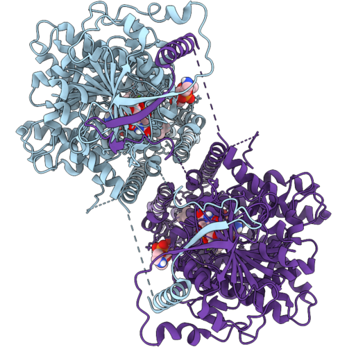
Candida Albicans Chitin Synthase 1 (Cachs1) Intermediate State (Da-4), Bound To Udp And Diynyl Arylamine (Da, Aka Ro-09-3143)
Organism: Candida albicans
Method: ELECTRON MICROSCOPY Resolution:3.10 Å Release Date: 2026-10-14 Classification: MEMBRANE PROTEIN Ligands: PEE, MG, A1DEH, UDP |
Organism: Candida albicans
Method: ELECTRON MICROSCOPY
Release Date: 2026-10-14
Ligands: PEE, MG, A1DEH, UDP
 |
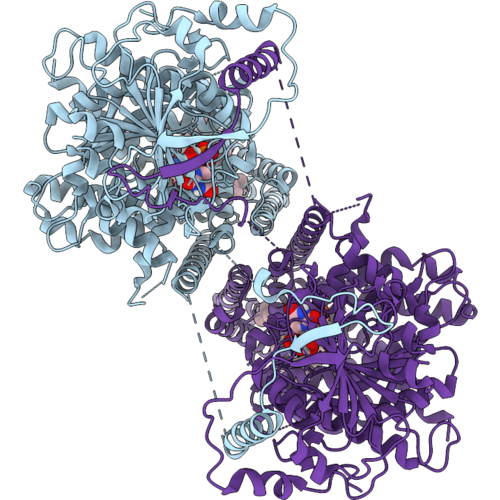
Candida Albicans Chitin Synthase 1 (Cachs1) Intermediate State (Da-5), Bound To Udp And Diynyl Arylamine (Da, Aka Ro-09-3143)
Organism: Candida albicans
Method: ELECTRON MICROSCOPY Resolution:2.85 Å Release Date: 2026-10-14 Classification: MEMBRANE PROTEIN Ligands: PEE, MG, A1DEH, UDP |
Organism: Candida albicans
Method: ELECTRON MICROSCOPY
Release Date: 2026-10-14
Ligands: PEE, MG, A1DEH, UDP
 |
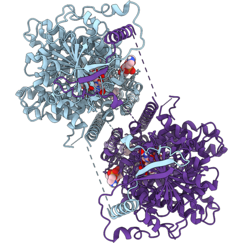
Candida Albicans Chitin Synthase 1 (Cachs1) Intermediate State (Da-6), Bound To Udp And Diynyl Arylamine (Da, Aka Ro-09-3143)
Organism: Candida albicans
Method: ELECTRON MICROSCOPY Resolution:3.29 Å Release Date: 2026-10-14 Classification: MEMBRANE PROTEIN Ligands: A1DEH, PEE, UDP |
Organism: Candida albicans
Method: ELECTRON MICROSCOPY
Release Date: 2026-10-14
Ligands: A1DEH, PEE, UDP
 |
Candida Albicans Chitin Synthase 1 (Cachs1) Intermediate State (Da-7), Bound To Udp And Diynyl Arylamine (Da, Aka Ro-09-3143)
Organism: Candida albicans
Method: ELECTRON MICROSCOPY Resolution:3.10 Å Release Date: 2026-10-14 Classification: MEMBRANE PROTEIN Ligands: PEE, A1DEH, UDP |
Organism: Candida albicans
Method: ELECTRON MICROSCOPY
Release Date: 2026-10-14
Ligands: PEE, A1DEH, UDP
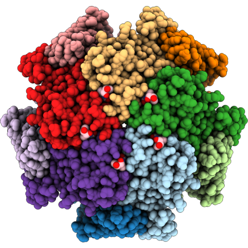 |
Cryo-Em Structure Of Gtp Cyclohydrolase I From Candida Albicans Bound To 7-Deaza Gtp And Bh2 At 1.96 A
Organism: Candida albicans
Method: ELECTRON MICROSCOPY Resolution:1.95 Å Release Date: 2026-09-09 Classification: ANTIFUNGAL PROTEIN Ligands: HBI, ZN, QBQ |
Organism: Candida albicans
Method: ELECTRON MICROSCOPY
Release Date: 2026-09-09
Ligands: HBI, ZN, QBQ
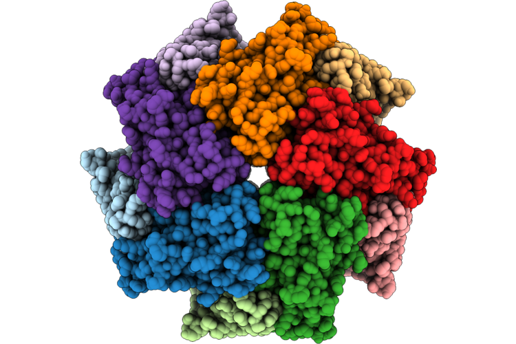 |
Cryo-Em Structure Of Gtp Cyclohydrolase I From Candida Albicans Bound To 8-Oxo Gtp At 1.74 A
Organism: Candida albicans
Method: ELECTRON MICROSCOPY Resolution:1.74 Å Release Date: 2026-08-26 Classification: ANTIFUNGAL PROTEIN Ligands: ZN, 8GT |
Organism: Candida albicans
Method: ELECTRON MICROSCOPY
Release Date: 2026-08-26
Ligands: ZN, 8GT
 |
Cryo-Em Structure Of Gtp Cyclohydrolase I From Candida Albicans At 2.6 A
Organism: Candida albicans
Method: ELECTRON MICROSCOPY Resolution:2.62 Å Release Date: 2026-08-19 Classification: ANTIFUNGAL PROTEIN Ligands: ZN |
Organism: Candida albicans
Method: ELECTRON MICROSCOPY
Release Date: 2026-08-19
Ligands: ZN
 |
The Structure Of Phosphoglucose Isomerase In Complex With Deoxynojirimycin-6-Phosphate And A Fragment
Organism: Candida albicans
Method: X-RAY DIFFRACTION Resolution:1.70 Å Release Date: 2026-08-12 Classification: ISOMERASE Ligands: IRU, A1JCV, CL |
Organism: Candida albicans
Method: X-RAY DIFFRACTION
Release Date: 2026-08-12
Ligands: IRU, A1JCV, CL
 |
The Structure Of Candida Albicans Phosphoglucose Isomerase In Complex With 1-Deoxynojirimycin-6-Phosphate
Organism: Candida albicans
Method: X-RAY DIFFRACTION Resolution:1.71 Å Release Date: 2026-06-17 Classification: ISOMERASE Ligands: A1JCV, NA |
Organism: Candida albicans
Method: X-RAY DIFFRACTION
Release Date: 2026-06-17
Ligands: A1JCV, NA
 |
Crystal Structure Of Candida Albicans Eukaryotic Translation Initiation Factor 5A
Organism: Candida albicans
Method: X-RAY DIFFRACTION Resolution:2.60 Å Release Date: 2026-05-27 Classification: TRANSLATION |
Organism: Candida albicans
Method: X-RAY DIFFRACTION
Release Date: 2026-05-27
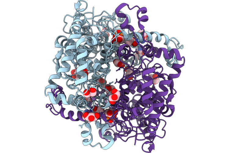 |
The Structure Of Capgi In Complex With Fragments
Organism: Candida albicans
Method: X-RAY DIFFRACTION Resolution:1.50 Å Release Date: 2026-05-20 Classification: ISOMERASE Ligands: GOL, A1JCW, A1JCV, CL, NA |
Organism: Candida albicans
Method: X-RAY DIFFRACTION
Release Date: 2026-05-20
Ligands: GOL, A1JCW, A1JCV, CL, NA
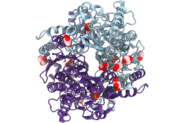 |
The Structure Of Capgi In Complex With Fragments
Organism: Candida albicans
Method: X-RAY DIFFRACTION Resolution:1.50 Å Release Date: 2026-05-20 Classification: ISOMERASE Ligands: GOL, A1JCV, CL, XKD |
Organism: Candida albicans
Method: X-RAY DIFFRACTION
Release Date: 2026-05-20
Ligands: GOL, A1JCV, CL, XKD