
Search Count: 29,001
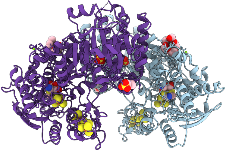 |
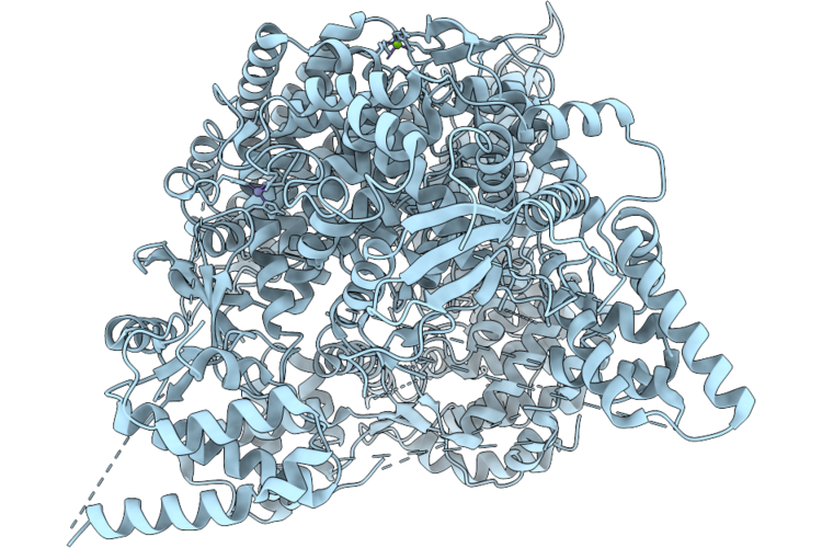
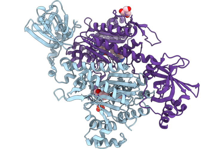
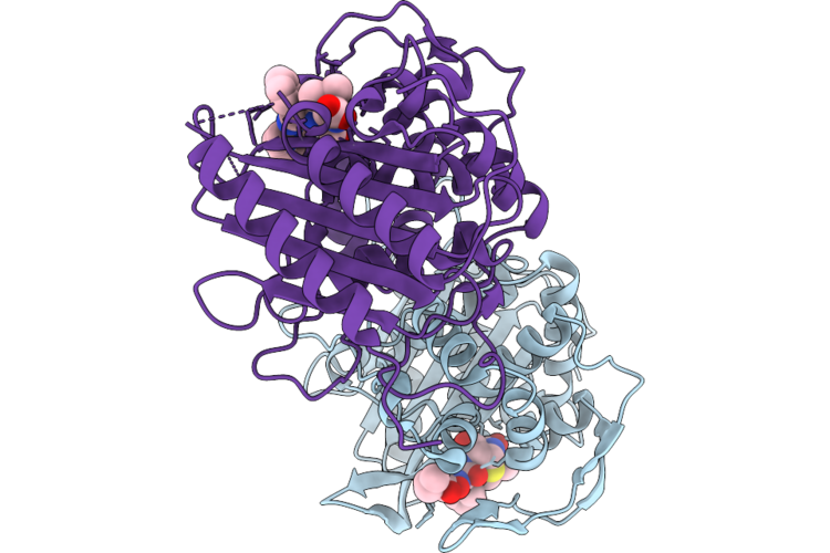
Crystal Structure Of [Fefe]-Hydrogenase Cba5H From Clostridium Beijerinckii In Hinact State
Organism: Clostridium beijerinckii
Method: X-RAY DIFFRACTION Resolution:1.96 Å Release Date: 2026-06-24 Classification: OXIDOREDUCTASE Ligands: 402, SF4, EPE, 1PE, ZN, MG, SO4 |
 |
Organism: Streptomyces sp. ajs327
Method: X-RAY DIFFRACTION Resolution:1.93 Å Release Date: 2026-06-17 Classification: FLAVOPROTEIN Ligands: FAD, CA, A1CGB |
 |
Organism: Streptomyces sp. ajs327
Method: X-RAY DIFFRACTION Resolution:1.56 Å Release Date: 2026-06-17 Classification: FLAVOPROTEIN Ligands: FAD, CA |
 |
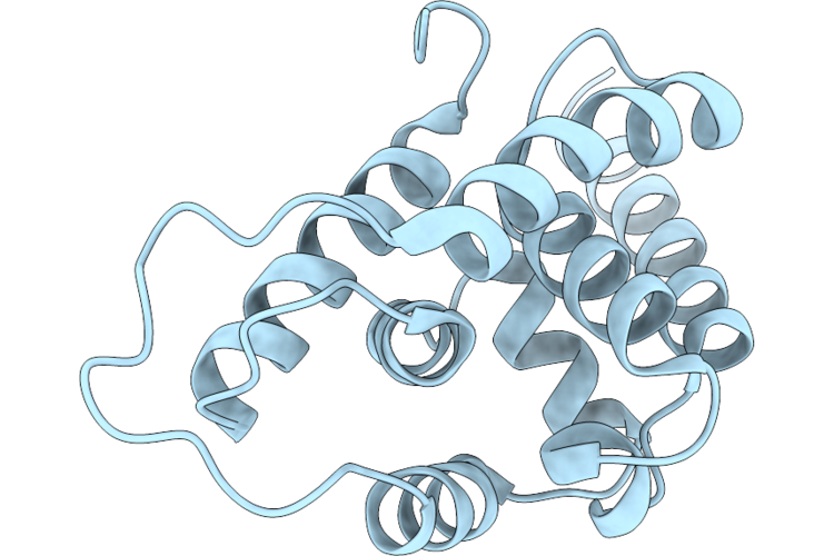
Crystal Structure Of Conserved Hypothetical Protein From Stenotrophomonas Maltophilia (Strain K279A)
Organism: Stenotrophomonas maltophilia k279a
Method: X-RAY DIFFRACTION Resolution:1.85 Å Release Date: 2026-06-17 Classification: UNKNOWN FUNCTION Ligands: SO4, PGE, CL, EDO, EPE |
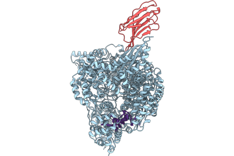 |
Structure Of Crimean Congo Hemorrhagic Fever Virus (Cchfv) L Protein Bound To 5' Vrna And Nanobody 20096.
Organism: Lama glama, Crimean-congo hemorrhagic fever virus strain ibar10200
Method: ELECTRON MICROSCOPY Release Date: 2026-06-17 Classification: VIRAL PROTEIN Ligands: ZN, MG |
 |
Organism: Crimean-congo hemorrhagic fever virus strain ibar10200
Method: ELECTRON MICROSCOPY Release Date: 2026-06-17 Classification: VIRAL PROTEIN Ligands: ZN, MG |
 |
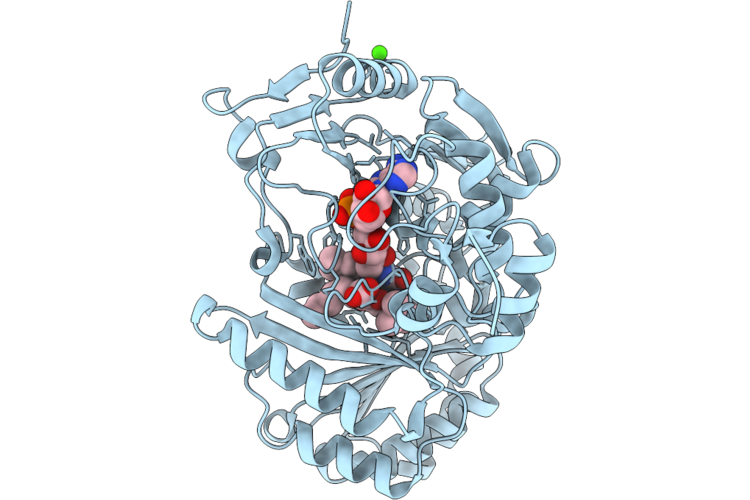
Hosa Transcriptional Regulator From Enteropathogenic Escherichia Coli O127:H6 (Strain E2348/69) Bound With 4-Hydroxy Benzoic Acid - L2' Conformation At 1.41 Angstrom Resolution
Organism: Escherichia coli o127:h6 str. e2348/69
Method: X-RAY DIFFRACTION Resolution:1.41 Å Release Date: 2026-06-17 Classification: DNA BINDING PROTEIN Ligands: PHB, PEG, CL, NA |
 |
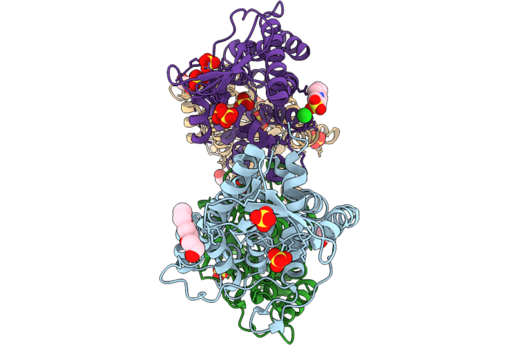
Crystal Structure Of Substrate Binding Protein (Taxi-Trap) In Complex With L-Glutamate From Bordetella Pertussis
Organism: Bordetella pertussis
Method: X-RAY DIFFRACTION Resolution:2.01 Å Release Date: 2026-06-10 Classification: SUBSTRATE BINDING PROTEIN Ligands: GLU |
 |
Organism: Streptomyces thermoviolaceus
Method: X-RAY DIFFRACTION Resolution:1.41 Å Release Date: 2026-06-10 Classification: DNA BINDING PROTEIN |
 |
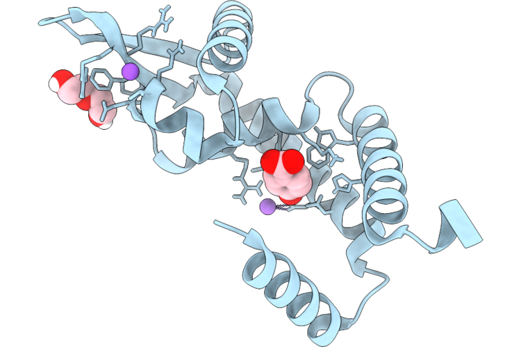
Streptomyces Thermoviolaceus Clpc2 C-Terminal Domain With Bound Phosphoarginine
Organism: Streptomyces thermoviolaceus
Method: X-RAY DIFFRACTION Resolution:1.30 Å Release Date: 2026-06-10 Classification: DNA BINDING PROTEIN Ligands: EDO, PEG, RPI |
 |
Streptomyces Thermoviolaceus Clpc2 C-Terminal Domain With Bound Cyclomarin A
Organism: Streptomyces thermoviolaceus, Streptomyces sp.
Method: X-RAY DIFFRACTION Resolution:1.55 Å Release Date: 2026-06-10 Classification: DNA BINDING PROTEIN Ligands: EDO |
 |
Organism: Cryptosporidium parvum iowa ii
Method: X-RAY DIFFRACTION Resolution:1.84 Å Release Date: 2026-06-10 Classification: LIGASE Ligands: LYS, A1JCS, SO4, TRS |
 |
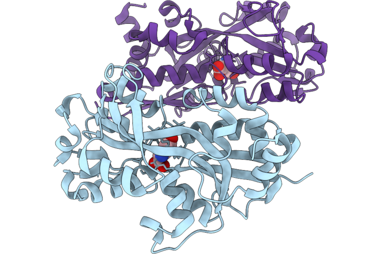
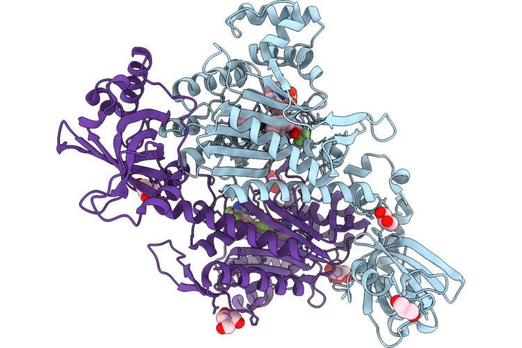
Crystal Structure Of Lysyl-Trna Synthetase From Cryptosporidium Parvum Complexed With L-Lysine And Inhibitor Ddd01887015
Organism: Cryptosporidium parvum iowa ii
Method: X-RAY DIFFRACTION Resolution:1.90 Å Release Date: 2026-06-10 Classification: LIGASE Ligands: A1JCT, EDO, TRS, GOL, LYS, SO4 |
 |
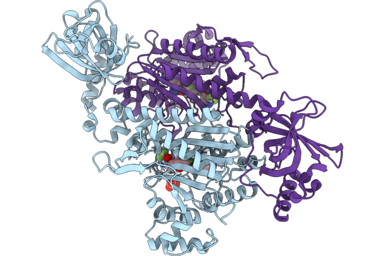
Crystal Structure Of Lysyl-Trna Synthetase From Cryptosporidium Parvum Complexed With L-Lysine And Inhibitor Ddd01932549
Organism: Cryptosporidium parvum iowa ii
Method: X-RAY DIFFRACTION Resolution:2.30 Å Release Date: 2026-06-10 Classification: LIGASE Ligands: LYS, A1JC7, SO4 |
 |
Crystal Structure Of Lysyl-Trna Synthetase From Cryptosporidium Parvum / Plasmodium Falciparum Chimera Complexed With L-Lysine And Inhibitor Ddd02174286
Organism: Cryptosporidium parvum iowa ii
Method: X-RAY DIFFRACTION Resolution:1.90 Å Release Date: 2026-06-10 Classification: LIGASE Ligands: LYS, A1JC4, GOL, SO4, TRS |
 |
Crystal Structure Of Lysyl-Trna Synthetase From Cryptosporidium Parvum Complexed With L-Lysine And Inhibitor Ddd01827593
Organism: Cryptosporidium parvum iowa ii
Method: X-RAY DIFFRACTION Resolution:1.50 Å Release Date: 2026-06-10 Classification: LIGASE Ligands: LYS, A1JC9, SO4 |
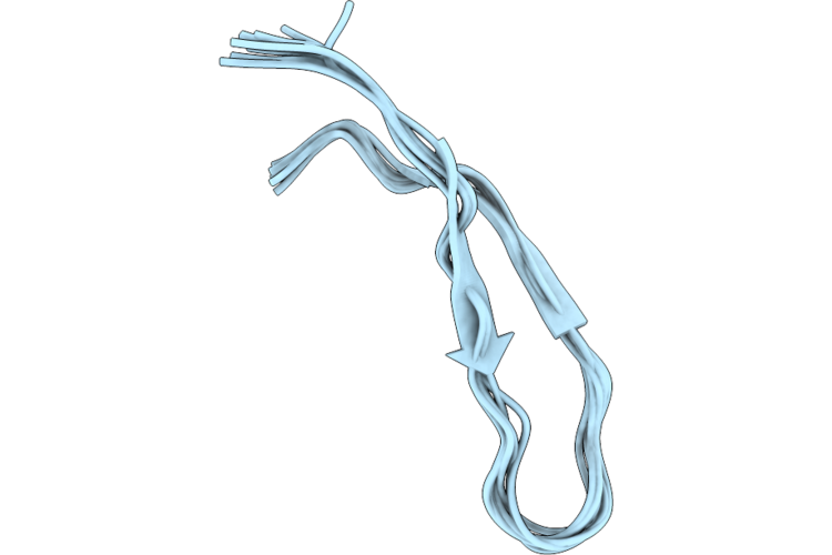 |
Organism: Abarenicola pacifica
Method: SOLUTION NMR Release Date: 2026-06-10 Classification: ANTIBIOTIC |
 |
Organism: Abarenicola pacifica
Method: SOLUTION NMR Release Date: 2026-06-10 Classification: ANTIBIOTIC |
 |
Crystal Structure Of The Transpeptidase Domain Of Pbp2 From Neisseria Gonorrhoeae Strain Fa19 Acylated By Piperacillin
Organism: Neisseria gonorrhoeae fa19
Method: X-RAY DIFFRACTION Resolution:3.15 Å Release Date: 2026-06-03 Classification: LIGASE Ligands: JPP |
 |
Organism: Dianlovirus menglaense, Homo sapiens
Method: ELECTRON MICROSCOPY Resolution:3.47 Å Release Date: 2026-06-03 Classification: VIRAL PROTEIN Ligands: NAG |