

Search Count: 17
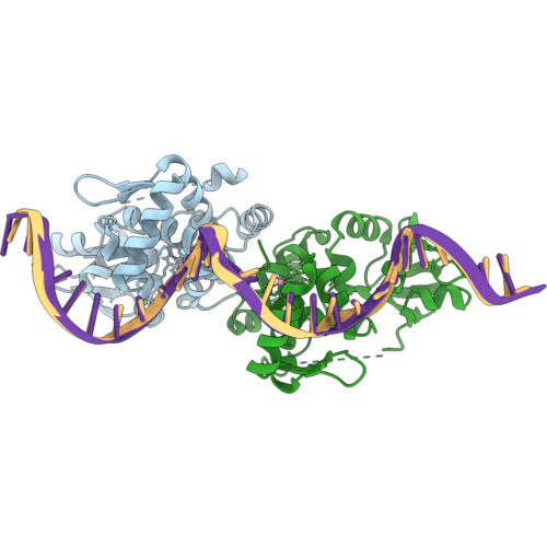 |
Dtxr-Like Iron-Dependent Regulator Ider Complexed With Cobalt And A Mutated Consensus Dna-Binding Sequence
Organism: Saccharopolyspora erythraea nrrl 2338, Synthetic construct
Method: X-RAY DIFFRACTION Resolution:2.26 Å Release Date: 2026-10-07 Classification: TRANSCRIPTION Ligands: CO |
Organism: Saccharopolyspora erythraea nrrl 2338, Synthetic construct
Method: X-RAY DIFFRACTION
Release Date: 2026-10-07
Ligands: CO
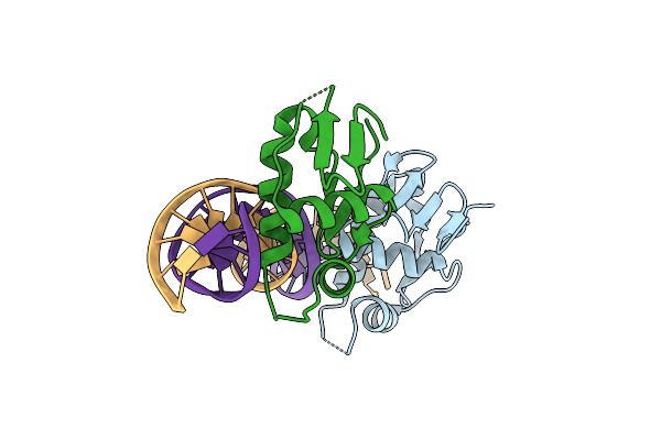 |
Co-Crystal Structure Of C-Terminal Dna Binding Domain Of Saccharopolyspora Erythraea Glnr In Complex With Its Cognate Promoter Dna
Organism: Saccharopolyspora erythraea nrrl 2338
Method: X-RAY DIFFRACTION Resolution:2.33 Å Release Date: 2023-11-22 Classification: TRANSCRIPTION/DNA |
Organism: Saccharopolyspora erythraea nrrl 2338
Method: X-RAY DIFFRACTION
Release Date: 2023-11-22
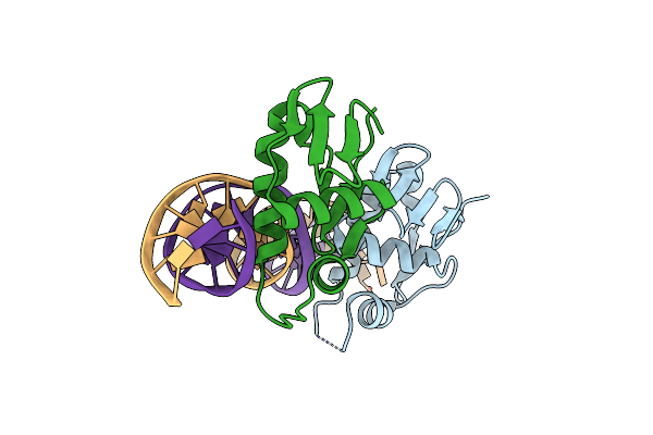 |
Co-Crystal Structure Of The C Terminal Dna Binding Domain Of Saccharopolyspora Erythraea Glnr In Complex With Its Conserved Promoter Dna In 2.95 Angstrom Resolution
Organism: Saccharopolyspora erythraea nrrl 2338
Method: X-RAY DIFFRACTION Resolution:2.95 Å Release Date: 2023-06-07 Classification: TRANSFERASE/DNA |
Organism: Saccharopolyspora erythraea nrrl 2338
Method: X-RAY DIFFRACTION
Release Date: 2023-06-07
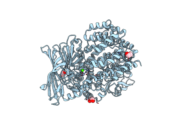 |
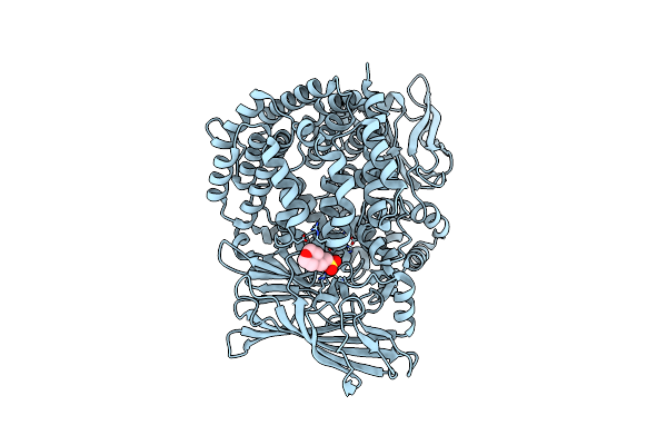
Crystal Structure Of The Lanthipeptide Zinc-Metallopeptidase Eryp From Saccharopolyspora Erythraea In Closed State
Organism: Saccharopolyspora erythraea (strain atcc 11635 / dsm 40517 / jcm 4748 / nbrc 13426 / ncimb 8594 / nrrl 2338)
Method: X-RAY DIFFRACTION Resolution:1.90 Å Release Date: 2022-05-11 Classification: HYDROLASE Ligands: ZN, CA, GOL, ACT |
Organism: Saccharopolyspora erythraea (strain atcc 11635 / dsm 40517 / jcm 4748 / nbrc 13426 / ncimb 8594 / nrrl 2338)
Method: X-RAY DIFFRACTION
Release Date: 2022-05-11
Ligands: ZN, CA, GOL, ACT
 |
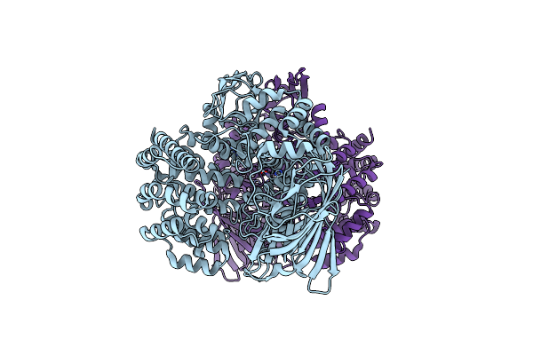
Crystal Structure Of The Lanthipeptide Zinc-Metallopeptidase Eryp Mutant E802R From Saccharopolyspora Erythraea
Organism: Saccharopolyspora erythraea (strain atcc 11635 / dsm 40517 / jcm 4748 / nbrc 13426 / ncimb 8594 / nrrl 2338)
Method: X-RAY DIFFRACTION Resolution:1.77 Å Release Date: 2022-05-11 Classification: HYDROLASE Ligands: ZN, MES |
 |
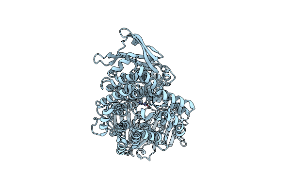
Crystal Structure Of The Lanthipeptide Zinc-Metallopeptidase Eryp From Saccharopolyspora Erythraea In Intermediate State
Organism: Saccharopolyspora erythraea (strain atcc 11635 / dsm 40517 / jcm 4748 / nbrc 13426 / ncimb 8594 / nrrl 2338)
Method: X-RAY DIFFRACTION Resolution:1.80 Å Release Date: 2022-05-11 Classification: HYDROLASE Ligands: ZN |
 |
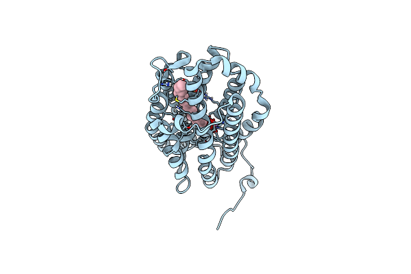
Crystal Structure Of The Lanthipeptide Zinc-Metallopeptidase Eryp From Saccharopolyspora Erythraea In Open State
Organism: Saccharopolyspora erythraea (strain atcc 11635 / dsm 40517 / jcm 4748 / nbrc 13426 / ncimb 8594 / nrrl 2338)
Method: X-RAY DIFFRACTION Resolution:2.67 Å Release Date: 2022-05-11 Classification: HYDROLASE Ligands: ZN |
 |
Crystal Structure Of R2-Like Ligand-Binding Oxidase From Saccharopolyspora Erythraea
Organism: Saccharopolyspora erythraea (strain atcc 11635 / dsm 40517 / jcm 4748 / nbrc 13426 / ncimb 8594 / nrrl 2338)
Method: X-RAY DIFFRACTION Resolution:1.38 Å Release Date: 2022-04-06 Classification: OXIDOREDUCTASE Ligands: MN3, FE, PLM, SO4 |
Organism: Saccharopolyspora erythraea (strain atcc 11635 / dsm 40517 / jcm 4748 / nbrc 13426 / ncimb 8594 / nrrl 2338)
Method: X-RAY DIFFRACTION
Release Date: 2022-04-06
Ligands: MN3, FE, PLM, SO4
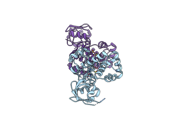 |
Dtxr-Like Iron-Dependent Regulator Ider Complexed With Cobalt
Organism: Saccharopolyspora erythraea (strain atcc 11635 / dsm 40517 / jcm 4748 / nbrc 13426 / ncimb 8594 / nrrl 2338)
Method: X-RAY DIFFRACTION Resolution:2.04 Å Release Date: 2021-03-03 Classification: TRANSCRIPTION Ligands: CO |
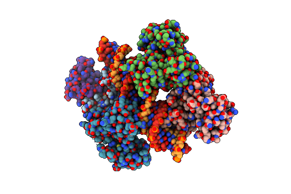 |
Dtxr-Like Iron-Dependent Regulator Ider Complexed With Cobalt And Its Consensus Dna-Binding Sequence
Organism: Saccharopolyspora erythraea (strain atcc 11635 / dsm 40517 / jcm 4748 / nbrc 13426 / ncimb 8594 / nrrl 2338), Synthetic construct
Method: X-RAY DIFFRACTION Resolution:2.12 Å Release Date: 2021-03-03 Classification: TRANSCRIPTION Ligands: CO |
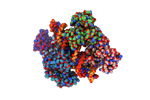 |
Dtxr-Like Iron-Dependent Regulator Ider Complexed With Iron And Its Consensus Dna-Binding Sequence
Organism: Saccharopolyspora erythraea (strain atcc 11635 / dsm 40517 / jcm 4748 / nbrc 13426 / ncimb 8594 / nrrl 2338), Synthetic construct
Method: X-RAY DIFFRACTION Resolution:2.18 Å Release Date: 2021-03-03 Classification: TRANSCRIPTION Ligands: FE2 |
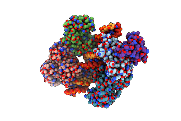 |
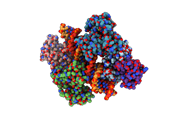
Dtxr-Like Iron-Dependent Regulator Ider Complexed With Cobalt And The Sace_2689 Promoter Dna-Binding Sequence
Organism: Saccharopolyspora erythraea (strain atcc 11635 / dsm 40517 / jcm 4748 / nbrc 13426 / ncimb 8594 / nrrl 2338), Synthetic construct
Method: X-RAY DIFFRACTION Resolution:2.15 Å Release Date: 2021-03-03 Classification: TRANSCRIPTION Ligands: CO |
 |
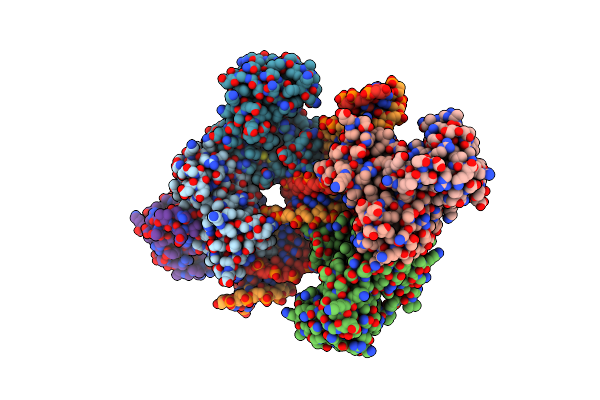
Dtxr-Like Iron-Dependent Regulator Ider (P39G Variant) Complexed With Cobalt And Its Consensus Dna-Binding Sequence
Organism: Saccharopolyspora erythraea (strain atcc 11635 / dsm 40517 / jcm 4748 / nbrc 13426 / ncimb 8594 / nrrl 2338), Synthetic construct
Method: X-RAY DIFFRACTION Resolution:2.05 Å Release Date: 2021-03-03 Classification: TRANSCRIPTION Ligands: CO |
 |
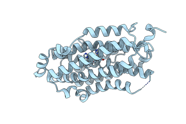
Dtxr-Like Iron-Dependent Regulator Ider (Q43A Variant) Complexed With Cobalt And Its Consensus Dna-Binding Sequence
Organism: Saccharopolyspora erythraea (strain atcc 11635 / dsm 40517 / jcm 4748 / nbrc 13426 / ncimb 8594 / nrrl 2338), Synthetic construct
Method: X-RAY DIFFRACTION Resolution:2.34 Å Release Date: 2021-03-03 Classification: TRANSCRIPTION Ligands: CO |
 |
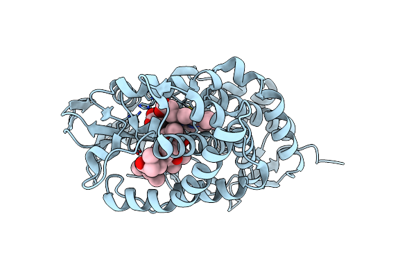
Crystal Structure Of Ribonucleotide Reductase R2 Subunit Solved By Serial Synchrotron Crystallography
Organism: Saccharopolyspora erythraea (strain atcc 11635 / dsm 40517 / jcm 4748 / nbrc 13426 / ncimb 8594 / nrrl 2338)
Method: X-RAY DIFFRACTION Resolution:2.40 Å Release Date: 2020-10-07 Classification: OXIDOREDUCTASE Ligands: MN3, FE |
 |
Structure Of A Mutant Of P450 Eryk In Complex With Erythromycin B.
Organism: Saccharopolyspora erythraea nrrl 2338
Method: X-RAY DIFFRACTION Resolution:2.00 Å Release Date: 2013-05-01 Classification: OXIDOREDUCTASE/ANTIBIOTIC Ligands: HEM, ERB |
Organism: Saccharopolyspora erythraea nrrl 2338
Method: X-RAY DIFFRACTION
Release Date: 2013-05-01
Ligands: HEM, ERB
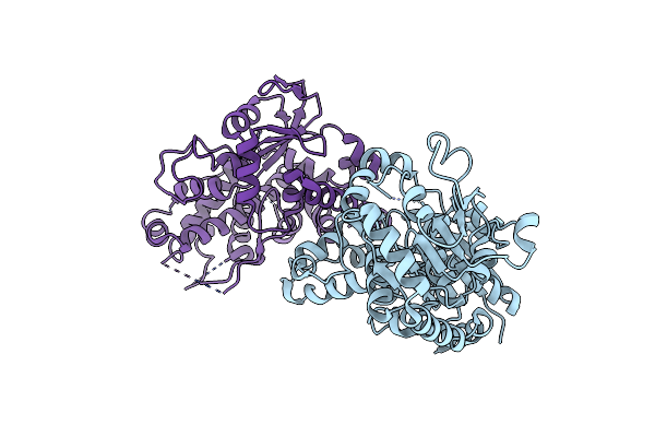 |
Structure Of The Glycosyltransferase Eryciii From The Erythromycin Biosynthetic Pathway, In Complex With Its Activating Partner, Erycii
Organism: Saccharopolyspora erythraea
Method: X-RAY DIFFRACTION Resolution:3.09 Å Release Date: 2011-11-09 Classification: TRANSFERASE |
Organism: Saccharopolyspora erythraea
Method: X-RAY DIFFRACTION
Release Date: 2011-11-09