
Search Count: 1,449
 |
Organism: Actinosynnema sp. ali-1.44
Method: X-RAY DIFFRACTION Resolution:2.60 Å Release Date: 2026-05-27 Classification: BIOSYNTHETIC PROTEIN |
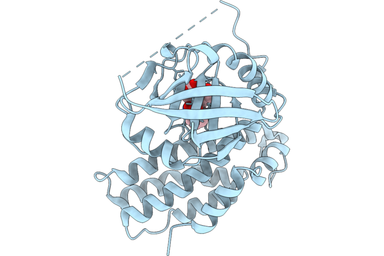 |
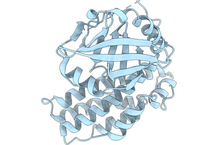
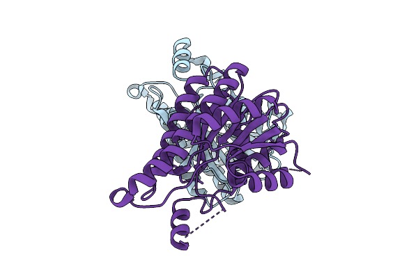
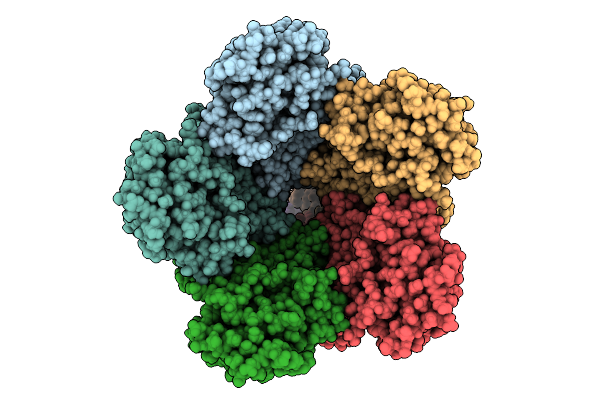
Crystal Structure Of Pictet-Spenglerase Askslb And The Compound Askslb With Iminium Ion Intermediate
Organism: Actinosynnema sp. ali-1.44
Method: X-RAY DIFFRACTION Resolution:1.70 Å Release Date: 2026-05-27 Classification: BIOSYNTHETIC PROTEIN Ligands: A1EQG |
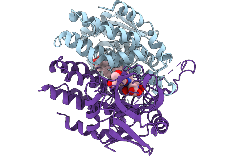 |
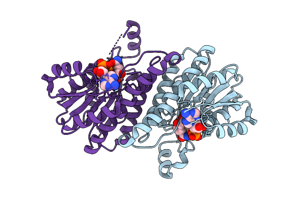
Pictet-Spenglerase Askslb In Complex With Product Of L-Trp And A-Ketoglutaric Acid
Organism: Actinosynnema sp. ali-1.44
Method: X-RAY DIFFRACTION Resolution:2.11 Å Release Date: 2026-05-27 Classification: BIOSYNTHETIC PROTEIN Ligands: 144, A1L46 |
 |
Organism: Human papillomavirus 16
Method: ELECTRON MICROSCOPY Resolution:1.90 Å Release Date: 2026-04-29 Classification: VIRUS Ligands: MG |
 |
Organism: Feline infectious peritonitis virus (strain 79-1146), Enterobacteria phage t6
Method: ELECTRON MICROSCOPY Resolution:2.78 Å Release Date: 2026-01-28 Classification: PROTEIN BINDING Ligands: NAG |
 |
Organism: Murid gammaherpesvirus 4
Method: ELECTRON MICROSCOPY Resolution:3.30 Å Release Date: 2026-01-21 Classification: VIRAL PROTEIN |
 |
Organism: Feline infectious peritonitis virus (strain 79-1146), Felis catus
Method: ELECTRON MICROSCOPY Release Date: 2026-01-07 Classification: PROTEIN BINDING Ligands: NAG |
 |
Crystal Structure Of L-2-Keto-3-Deoxypentonate 4-Dehydrogenase Bound To Nad(H)
Organism: Herbaspirillum huttiense
Method: X-RAY DIFFRACTION Resolution:2.48 Å Release Date: 2025-11-26 Classification: OXIDOREDUCTASE Ligands: NAD |
 |
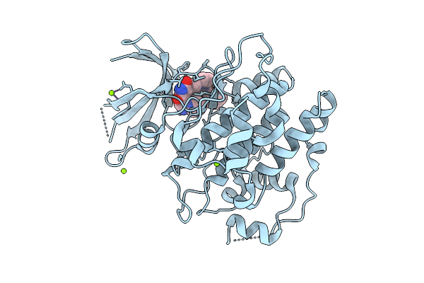
Toxoplasma Gondii Gsk3B Bound To Ly2090314 And Disulphide Bonded Through The C223 Residue
Organism: Toxoplasma gondii rh
Method: X-RAY DIFFRACTION Resolution:2.90 Å Release Date: 2025-10-22 Classification: TRANSFERASE Ligands: A1IYF, PO4 |
 |
Organism: Toxoplasma gondii rh
Method: X-RAY DIFFRACTION Resolution:2.10 Å Release Date: 2025-10-15 Classification: TRANSFERASE Ligands: A1IYF, MG |
 |
Crysral Structure Of 2-Keto-3-Deoxypentonate 4-Dehydrogenase From Herbaspirillum Huttiense (Apo Form)
Organism: Herbaspirillum huttiense
Method: X-RAY DIFFRACTION Resolution:2.27 Å Release Date: 2025-10-15 Classification: OXIDOREDUCTASE |
 |
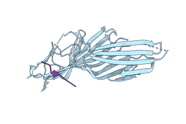
Crystal Structure Of The Mu2 Subunit Of The Clathrin-Adaptor Protein 2 (Ap2) Bound To Hpv16 E7(Residues 22-39)
Organism: Rattus norvegicus, Human papillomavirus 16
Method: X-RAY DIFFRACTION Resolution:3.70 Å Release Date: 2025-09-10 Classification: ENDOCYTOSIS |
 |
Crystal Structure Of The Mu2 Subunit Of The Clathrin-Adaptor Protein 2 (Ap2) Bound To Hpv16 E7(Residues 22-32; S31E And S32E)
Organism: Rattus norvegicus, Human papillomavirus 16
Method: X-RAY DIFFRACTION Resolution:3.20 Å Release Date: 2025-09-10 Classification: ENDOCYTOSIS |
 |
Crystal Structure Of The Mu2 Subunit Of The Clathrin-Adaptor Protein 2 (Ap2) Bound To Hpv16 E7(Residues 22-39; S31E And S32E)
Organism: Rattus norvegicus, Human papillomavirus 16
Method: X-RAY DIFFRACTION Resolution:3.30 Å Release Date: 2025-09-10 Classification: ENDOCYTOSIS |
 |
Organism: Hepatitis e virus (strain pakistan), Vicugna pacos
Method: X-RAY DIFFRACTION Resolution:2.20 Å Release Date: 2025-09-03 Classification: VIRAL PROTEIN/IMMUNE SYSTEM |
 |
Organism: Human papillomavirus 16, Homo sapiens
Method: ELECTRON MICROSCOPY Release Date: 2025-08-20 Classification: IMMUNE SYSTEM |
 |
Organism: Human papillomavirus 16, Homo sapiens
Method: ELECTRON MICROSCOPY Release Date: 2025-08-20 Classification: IMMUNE SYSTEM |
 |
Organism: Human papillomavirus 16, Homo sapiens
Method: ELECTRON MICROSCOPY Release Date: 2025-08-20 Classification: IMMUNE SYSTEM |
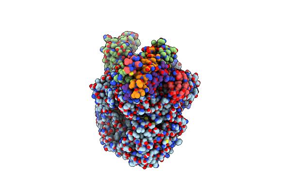 |
Cryo-Em Structure Of The Rna-Dependent Rna Polymerase Complex From Marburg Virus
Organism: Marburg virus - musoke, kenya, 1980, Escherichia coli k-12
Method: ELECTRON MICROSCOPY Resolution:2.70 Å Release Date: 2025-04-09 Classification: VIRAL PROTEIN Ligands: ZN |
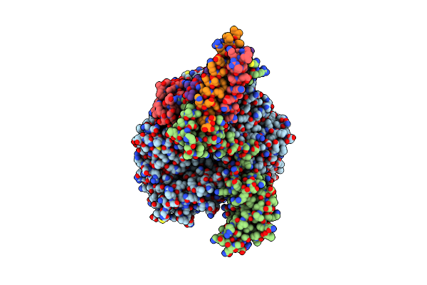 |
Cryo-Em Structure Of The Rna-Dependent Rna Polymerase Complex From Marburg Virus
Organism: Marburg virus - musoke, kenya, 1980, Escherichia coli k-12
Method: ELECTRON MICROSCOPY Resolution:2.84 Å Release Date: 2025-04-09 Classification: VIRAL PROTEIN Ligands: ZN |