
Search Count: 4,790
 |
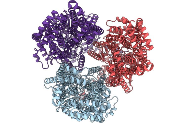
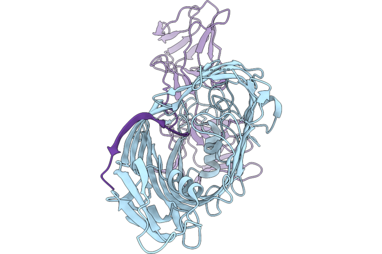
Cryo-Em Structure Of Mexjk From Pseudomonas Aeruginosa
Organism: Pseudomonas aeruginosa
Method: ELECTRON MICROSCOPY Resolution:2.57 Å Release Date: 2026-07-08 Classification: TRANSPORT PROTEIN Ligands: TCL |
 |
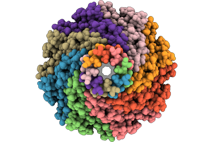
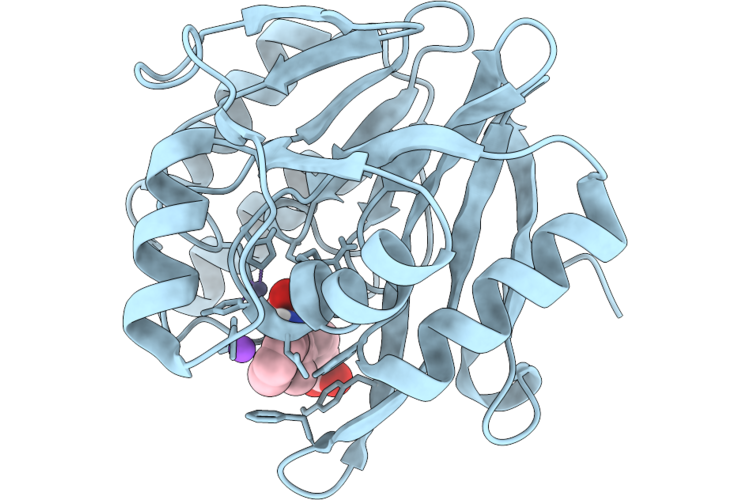
Crystal Structure Of Gamma Carbonic Anhydrase From Pseudomonas Aeruginosa (Pa3753)
Organism: Pseudomonas aeruginosa
Method: X-RAY DIFFRACTION Resolution:1.45 Å Release Date: 2026-07-08 Classification: LYASE Ligands: ZN, BME |
 |
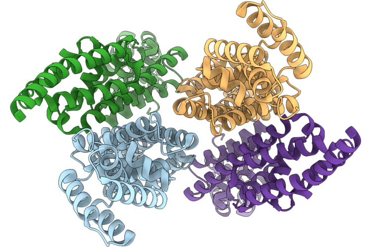
Cryo-Em Structure Of Mexk From Pseudomonas Aeruginosa
Organism: Pseudomonas aeruginosa
Method: ELECTRON MICROSCOPY Resolution:3.38 Å Release Date: 2026-07-08 Classification: TRANSPORT PROTEIN |
 |
Cryoem Struccture Of The Type Iii Secretion System Gatekeeper Protein Inve By Cryo-Electron Microscopy.
Organism: Salmonella enterica subsp. enterica serovar typhimurium str. lt2
Method: ELECTRON MICROSCOPY Release Date: 2026-07-01 Classification: CYTOSOLIC PROTEIN |
 |
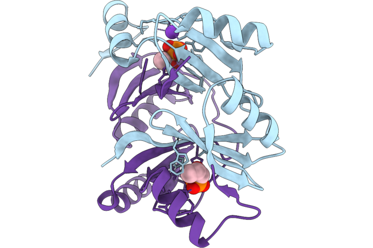
Crystal Structure Of Pdc-3 Beta-Lactamase Complexed With Boronic Acid Inhibitor Z2242032529
Organism: Pseudomonas aeruginosa
Method: X-RAY DIFFRACTION Resolution:1.98 Å Release Date: 2026-06-24 Classification: HYDROLASE/INHIBITOR Ligands: A1BI9 |
 |
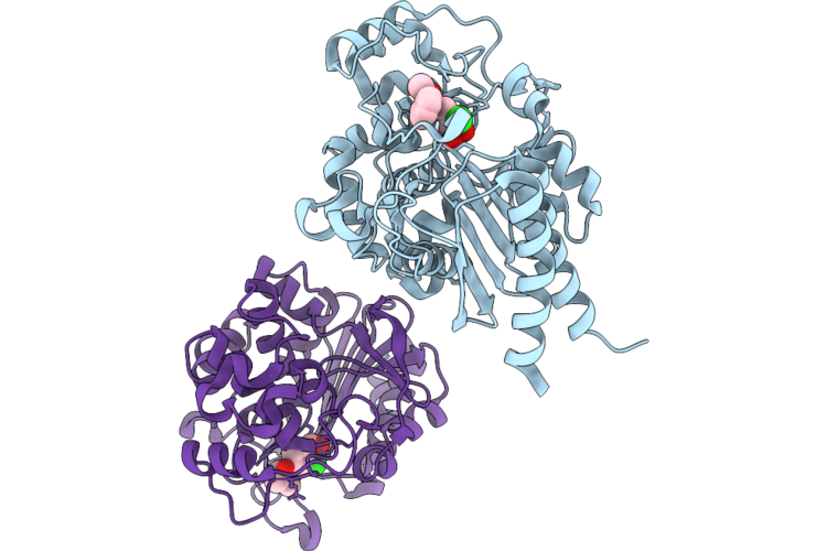
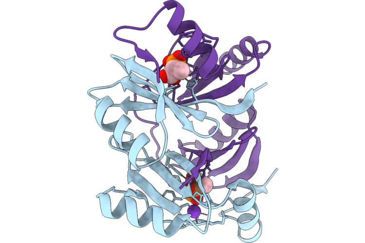
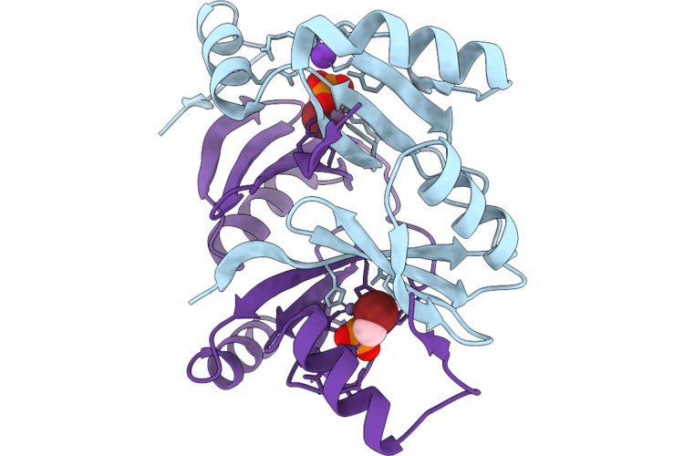
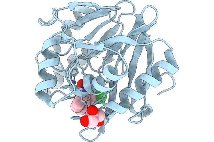
Crystal Structure Of Fosa From Pseudomonas Aeruginosa With Manganese And Phosphonoacetate
Organism: Pseudomonas aeruginosa
Method: X-RAY DIFFRACTION Resolution:1.41 Å Release Date: 2026-06-17 Classification: TRANSFERASE Ligands: MN, PAE, K |
 |
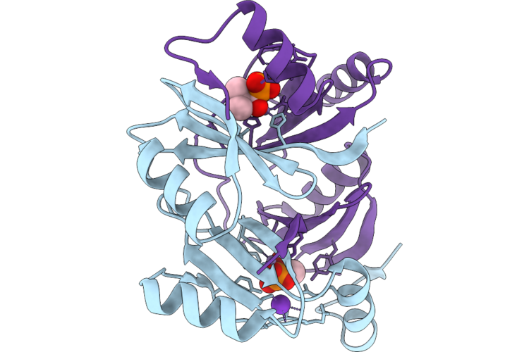
Crystal Structure Of Fosa From Pseudomonas Aeruginosa With Manganese And 2-Phosphonopropionic Acid
Organism: Pseudomonas aeruginosa
Method: X-RAY DIFFRACTION Resolution:1.80 Å Release Date: 2026-06-17 Classification: TRANSFERASE Ligands: MN, A1BHK, K |
 |
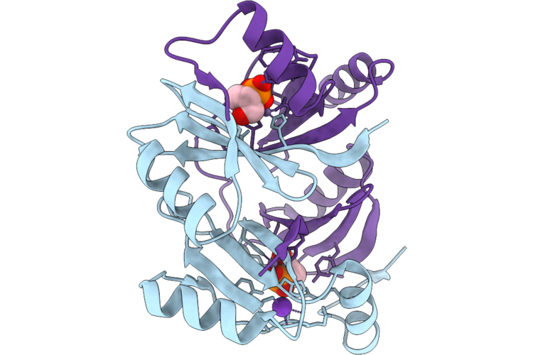
Crystal Structure Of Fosa From Pseudomonas Aeruginosa With Manganese And 1-Hydroxypropylphosphonic Acid
Organism: Pseudomonas aeruginosa
Method: X-RAY DIFFRACTION Resolution:1.47 Å Release Date: 2026-06-17 Classification: TRANSFERASE Ligands: MN, 1JJ, K |
 |
Crystal Structure Of Fosa From Pseudomonas Aeruginosa With Manganese And (1-Hydroxy-2-Methylpropyl)Phosphonic Acid
Organism: Pseudomonas aeruginosa
Method: X-RAY DIFFRACTION Resolution:1.77 Å Release Date: 2026-06-17 Classification: TRANSFERASE Ligands: MN, YS8, K |
 |
Crystal Structure Of Fosa From Pseudomonas Aeruginosa With Manganese And (1-Hydroxypropan-2-Yl)Phosphonic Acid
Organism: Pseudomonas aeruginosa
Method: X-RAY DIFFRACTION Resolution:1.59 Å Release Date: 2026-06-17 Classification: TRANSFERASE Ligands: MN, YRQ, K |
 |
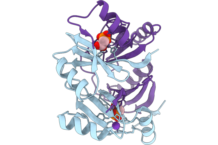
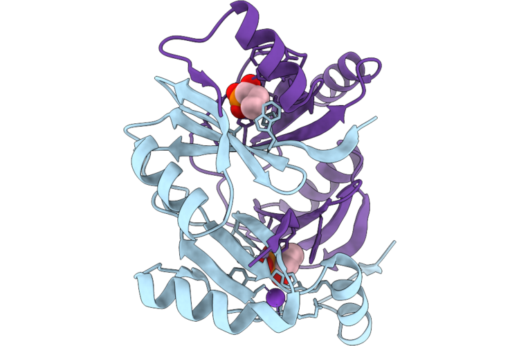
Crystal Structure Of Fosa From Pseudomonas Aeruginosa With Manganese And Fosfomycin
Organism: Pseudomonas aeruginosa
Method: X-RAY DIFFRACTION Resolution:1.80 Å Release Date: 2026-06-17 Classification: TRANSFERASE Ligands: FCN, MN, K |
 |
Crystal Structure Of Fosa From Pseudomonas Aeruginosa With Manganese And Chloromethylphosphonic Acid
Organism: Pseudomonas aeruginosa
Method: X-RAY DIFFRACTION Resolution:1.48 Å Release Date: 2026-06-17 Classification: TRANSFERASE/INHIBITOR Ligands: MN, A1CE8, K |
 |
Crystal Structure Of Fosa From Pseudomonas Aeruginosa And Bromomethyl Phosphonic Acid
Organism: Pseudomonas aeruginosa
Method: X-RAY DIFFRACTION Resolution:1.99 Å Release Date: 2026-06-17 Classification: TRANSFERASE/INHIBITOR Ligands: A1CE7, MN, K |
 |
Vgrg1 From Pseudomonas Aeruginosa (Pa0091)
Organism: Pseudomonas aeruginosa
Method: ELECTRON MICROSCOPY Release Date: 2026-06-10 Classification: TOXIN |
 |
Cryo-Em Structure Of Bam From P. Aeruginosa Pao1 In Complex With Pyocin L1
Organism: Pseudomonas aeruginosa, Pseudomonas aeruginosa pao1
Method: ELECTRON MICROSCOPY Release Date: 2026-06-10 Classification: MEMBRANE PROTEIN |
 |
Cryo-Em Structure Of Bam From P. Aeruginosa P28 In Complex With Pyocin L2
Organism: Pseudomonas aeruginosa, Pseudomonas aeruginosa 62
Method: ELECTRON MICROSCOPY Release Date: 2026-06-10 Classification: MEMBRANE PROTEIN |
 |
Psl Polysaccharide Related Protein Structures
Organism: Pseudomonas aeruginosa
Method: ELECTRON MICROSCOPY Resolution:3.06 Å Release Date: 2026-06-03 Classification: PROTEIN TRANSPORT |
 |
Psl Polysaccharide Related Protein Structures
Organism: Pseudomonas aeruginosa
Method: ELECTRON MICROSCOPY Release Date: 2026-06-03 Classification: PROTEIN TRANSPORT |
 |
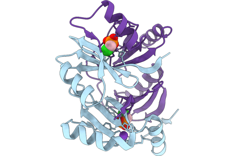
Crystal Structure Of The Metallo-Beta-Lactamase Vim-1 With 1476
Organism: Pseudomonas aeruginosa
Method: X-RAY DIFFRACTION Resolution:1.15 Å Release Date: 2026-05-27 Classification: HYDROLASE Ligands: GOL, A1JFP, ZN |
 |
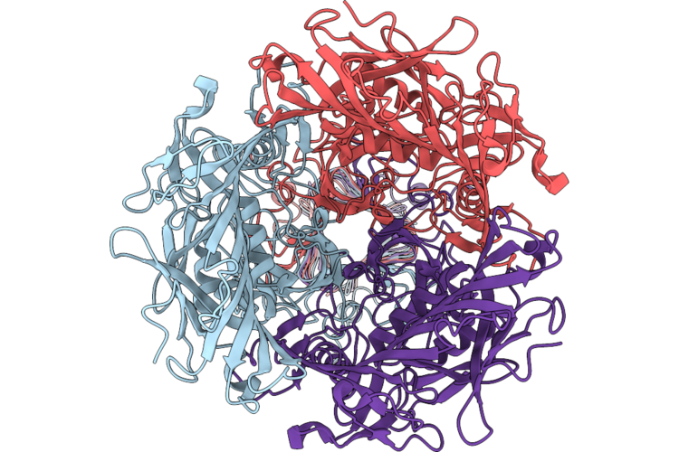
Crystal Structure Of The Metallo-Beta-Lactamase Vim-1 With 2495
Organism: Pseudomonas aeruginosa
Method: X-RAY DIFFRACTION Resolution:1.15 Å Release Date: 2026-05-27 Classification: HYDROLASE Ligands: A1JFL, ZN, NA |