
Search Count: 41,679
 |
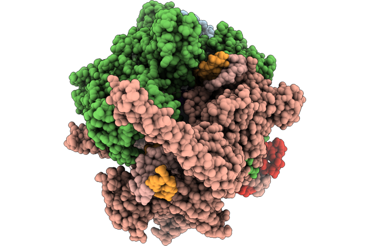
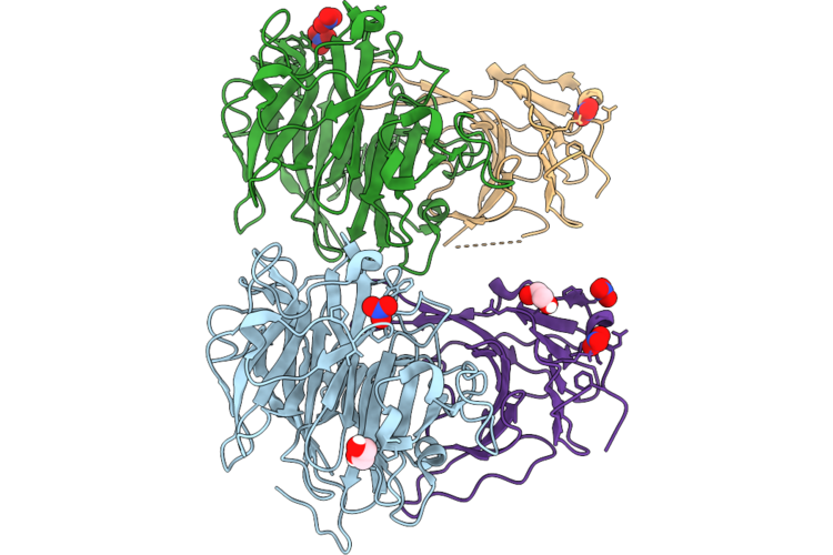
Closed Mtb-Ec: Cryo-Em Structure Of Mtb Rnap Elongation Complex (Substrate Loading Mimic) With A Closed Active Site (Closed Tl And Rh-Fl)
Organism: Mycobacterium tuberculosis, Escherichia coli
Method: ELECTRON MICROSCOPY Resolution:3.00 Å Release Date: 2026-06-24 Classification: TRANSCRIPTION Ligands: ZN, MG, GTP |
 |
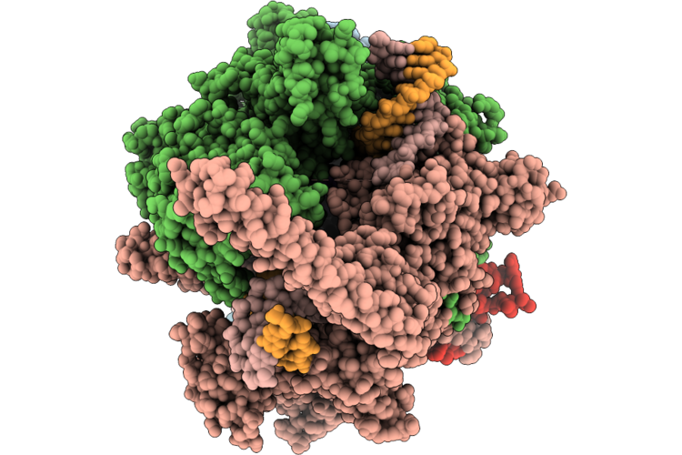
Open Mtb-Ec: Cryo-Em Structure Of Mtb Rnap Elongation Complex (Substrate Loading Mimic) With An Open Active Site (Open Tl And Rh-Fl)
Organism: Mycobacterium tuberculosis, Escherichia coli
Method: ELECTRON MICROSCOPY Resolution:3.30 Å Release Date: 2026-06-24 Classification: TRANSCRIPTION Ligands: ZN, MG |
 |
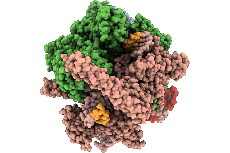
Semiclosed Mtb-Ec: Cryo-Em Structure Of Mtb Rnap Elongation Complex (Substrate Loading Mimic) With A Semiclosed Active Site (Closed Tl, Open Rh-Fl)
Organism: Mycobacterium tuberculosis, Escherichia coli
Method: ELECTRON MICROSCOPY Release Date: 2026-06-24 Classification: TRANSCRIPTION Ligands: GTP, ZN, MG |
 |
Aap-So2 Bound Open Mtb-Ec: Cryo-Em Structure Of Mtb Rnap Elongation Complex (Substrate Loading Mimic) With An Open Active Site (Open Tl And Rh-Fl)
Organism: Mycobacterium tuberculosis, Escherichia coli
Method: ELECTRON MICROSCOPY Resolution:3.00 Å Release Date: 2026-06-24 Classification: TRANSCRIPTION Ligands: GTP, A1BNV, MG, ZN |
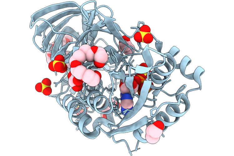 |
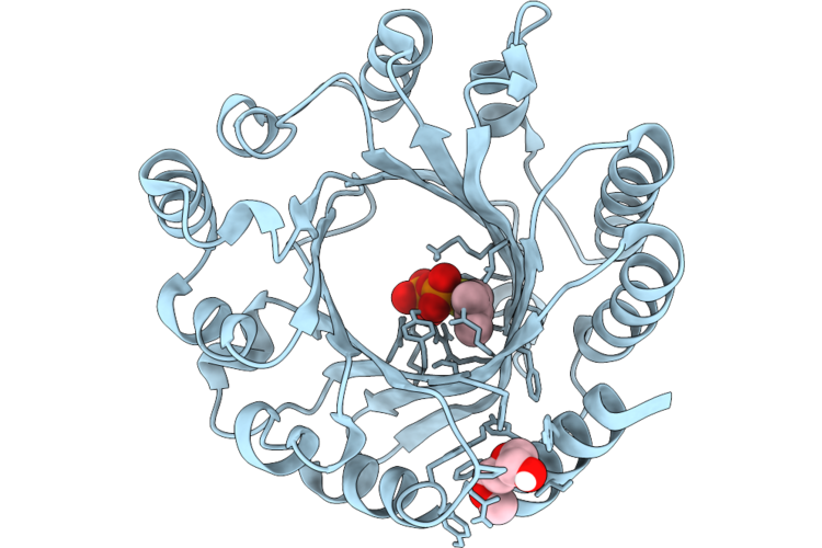
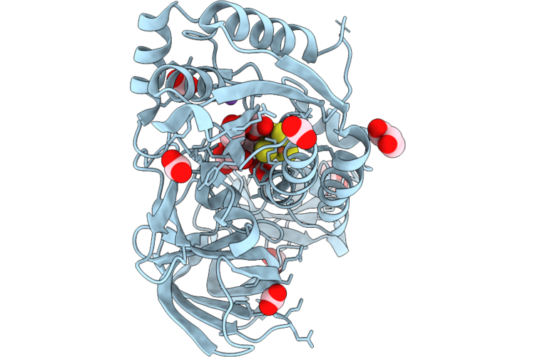
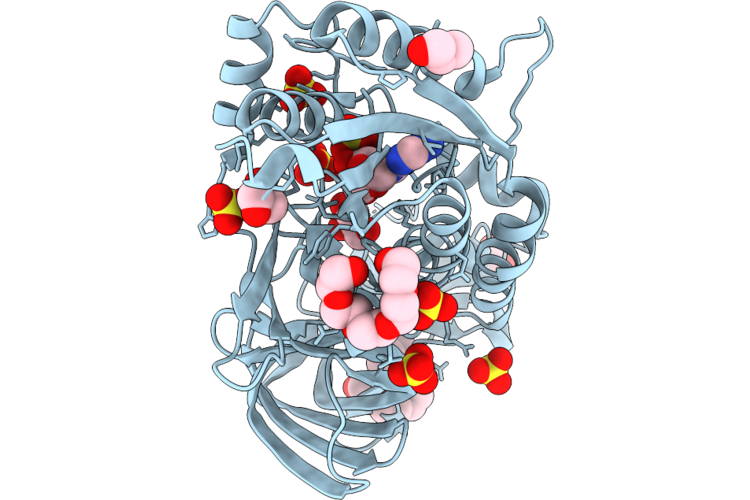
Crystal Structure Of Mnma From Streptococcus Pneumoniae In Complex With Atp
Organism: Streptococcus pneumoniae tigr4
Method: X-RAY DIFFRACTION Resolution:1.90 Å Release Date: 2026-06-24 Classification: TRANSFERASE Ligands: PEG, ATP, GOL, PG6, P6G, SO4 |
 |
Organism: Mycobacterium avium subsp. paratuberculosis
Method: X-RAY DIFFRACTION Resolution:1.65 Å Release Date: 2026-06-24 Classification: TOXIN,HYDROLASE Ligands: NO3, EDO |
 |
X-Ray Structure Of Leptospira Interrogans Histone Deacetylase 11 (Hdac11) In Complex With Cis-Dodec-5-Enoic Acid
Organism: Leptospira interrogans
Method: X-RAY DIFFRACTION Resolution:1.51 Å Release Date: 2026-06-24 Classification: HYDROLASE Ligands: GOL, A1JHJ, ZN, K, CL, NA, EDO |
 |
Crystal Structure Of Fgd2 From Mycobacterium Tuberculosis In Complex With Tew And Para Isopropylaniline (Fragment A6-11)
Organism: Mycobacterium tuberculosis
Method: X-RAY DIFFRACTION Resolution:1.89 Å Release Date: 2026-06-24 Classification: OXIDOREDUCTASE Ligands: IMD, ISO, TEW, MPD, CL, MES, BR |
 |
Organism: Mycolicibacterium smegmatis
Method: ELECTRON MICROSCOPY Resolution:3.26 Å Release Date: 2026-06-24 Classification: PROTEIN BINDING |
 |
Organism: Microcystis aeruginosa blcc-f108
Method: X-RAY DIFFRACTION Resolution:1.57 Å Release Date: 2026-06-24 Classification: TRANSFERASE Ligands: DST, PG0 |
 |
Organism: Paeonia ostii
Method: X-RAY DIFFRACTION Resolution:2.03 Å Release Date: 2026-06-24 Classification: TRANSFERASE Ligands: SAH |
 |
Organism: Paeonia ostii
Method: X-RAY DIFFRACTION Resolution:2.28 Å Release Date: 2026-06-24 Classification: TRANSFERASE Ligands: A1EVU, SAH |
 |
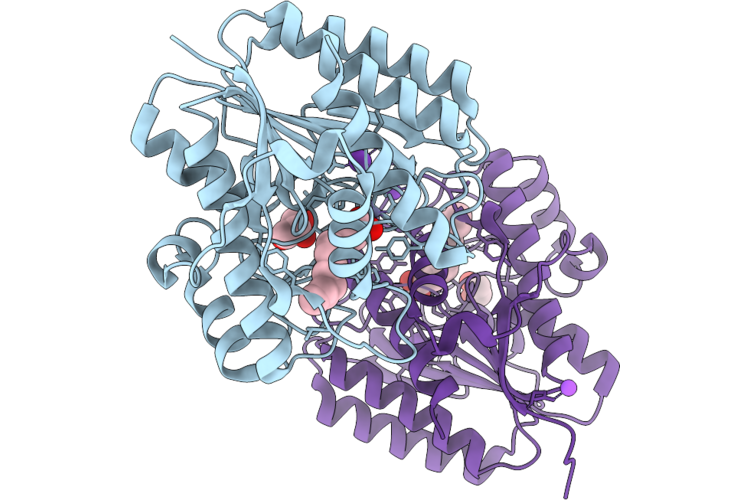
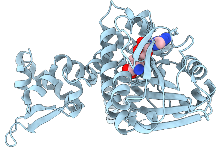
Crystal Structure Of Mnma D100C Mutant From Streptococcus Pneumoniae With [4Fe-4S] Cluster In Complex With Formate
Organism: Streptococcus pneumoniae tigr4
Method: X-RAY DIFFRACTION Resolution:1.92 Å Release Date: 2026-06-17 Classification: TRANSFERASE Ligands: SF4, FMT, GOL, NA |
 |
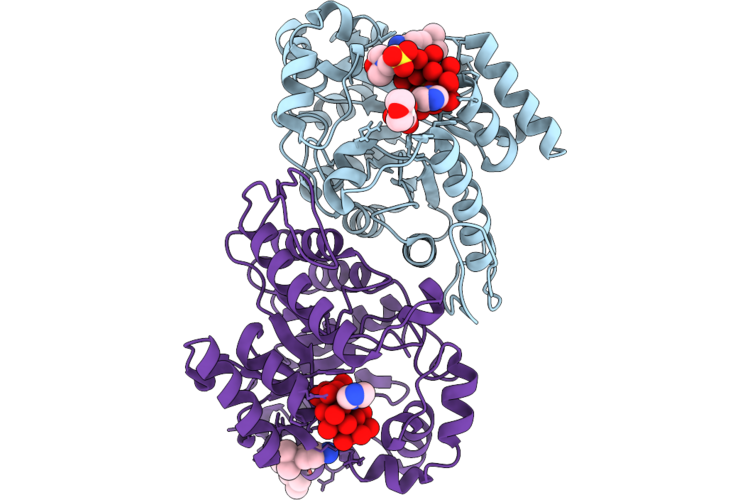
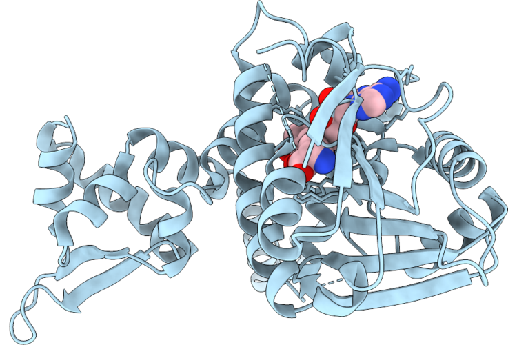
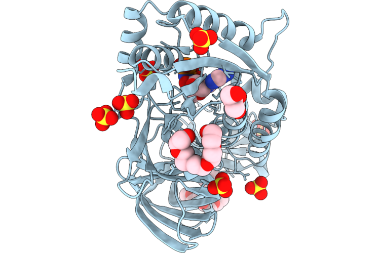
Crystal Structure Of Mnma From Streptococcus Pneumoniae In Complex With Amp
Organism: Streptococcus pneumoniae tigr4
Method: X-RAY DIFFRACTION Resolution:1.85 Å Release Date: 2026-06-17 Classification: TRANSFERASE Ligands: P6G, AMP, GOL, SO4 |
 |
Crystal Structure Of Mnma From Streptococcus Pneumoniae In Complex With Amp-Pnp
Organism: Streptococcus pneumoniae tigr4
Method: X-RAY DIFFRACTION Resolution:1.82 Å Release Date: 2026-06-17 Classification: TRANSFERASE Ligands: P6G, PG6, GOL, ANP, SO4 |
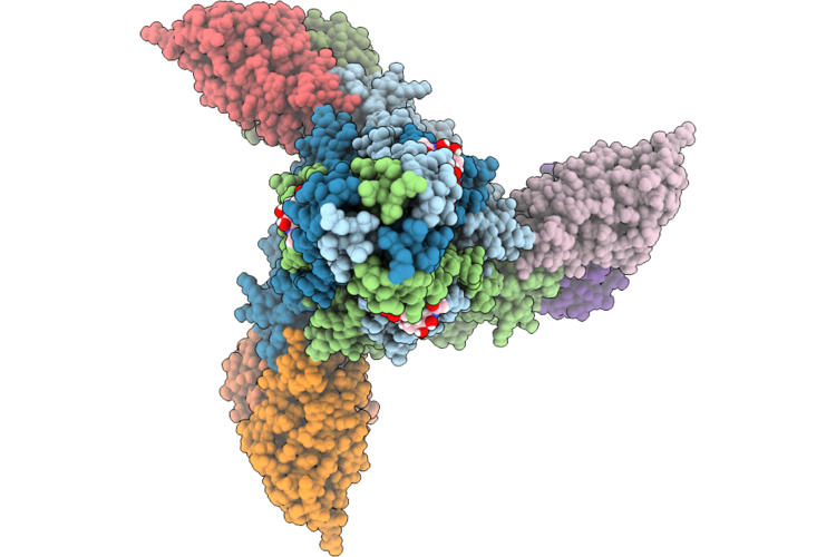 |
Cryo-Em Structure Of Vaccine Elicited Antibody 22F5 Bound To The Post-Fusion Conformation Of The Layv-F Glycoprotein
Organism: Langya virus, Mus musculus
Method: ELECTRON MICROSCOPY Release Date: 2026-06-17 Classification: IMMUNE SYSTEM/VIRAL PROTEIN Ligands: NAG |
 |
Organism: Rattus rattus, Homo sapiens, Escherichia coli, Synthetic construct, Human respiratory syncytial virus, Mus musculus
Method: ELECTRON MICROSCOPY Release Date: 2026-06-10 Classification: SIGNALING PROTEIN Ligands: UDP |
 |
Crystal Structure Of Monoalkyl Phthalate Hydrolase From Rhodococcus Sp. Eg-5
Organism: Rhodococcus sp. eg-5
Method: X-RAY DIFFRACTION Resolution:3.00 Å Release Date: 2026-06-10 Classification: HYDROLASE |
 |
Organism: Streptococcus pneumoniae r6
Method: X-RAY DIFFRACTION Resolution:2.80 Å Release Date: 2026-06-10 Classification: CELL CYCLE |
 |
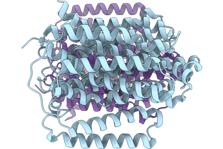
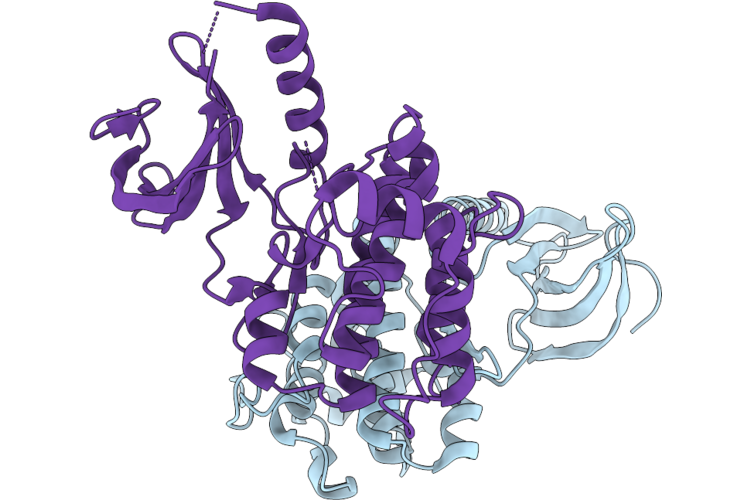
Streptococcus Pneumoniae Stkp Catalytic Domain T167E/T169E Double Mutant In Complex With Amp-Pnp And Mn2+
Organism: Streptococcus pneumoniae r6
Method: X-RAY DIFFRACTION Resolution:1.60 Å Release Date: 2026-06-10 Classification: CELL CYCLE Ligands: ANP, MN |