
Search Count: 160
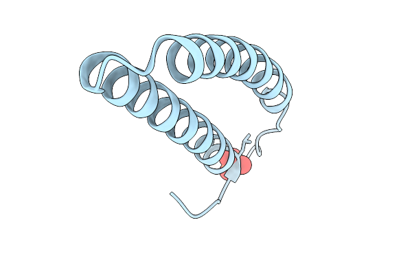 |
Crystal Structure Of A Escherichia Phage Upec07 Protein
Organism: Escherichia phage t4
Method: X-RAY DIFFRACTION Resolution:2.56 Å Release Date: 2026-03-04 Classification: ANTIMICROBIAL PROTEIN Ligands: SO4 |
Organism: Escherichia phage t4
Method: X-RAY DIFFRACTION
Release Date: 2026-03-04
Ligands: SO4
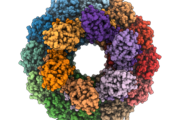 |
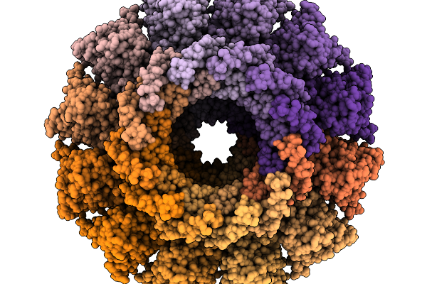
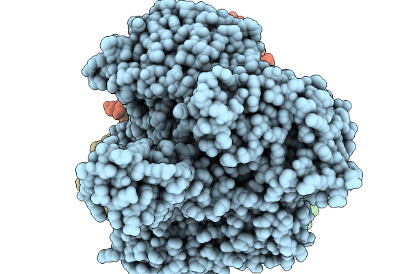
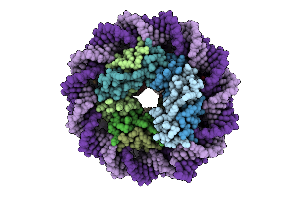
Structure Of Bacteriophage T4 Neck Protein Gp13 And Gp14 Assembled In Vitro In C6 Symmetry
Organism: Escherichia phage t4
Method: ELECTRON MICROSCOPY Release Date: 2026-02-18 Classification: VIRAL PROTEIN |
Organism: Escherichia phage t4
Method: ELECTRON MICROSCOPY
Release Date: 2026-02-18
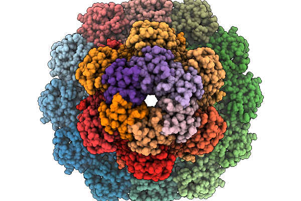 |
Structure Of Bacteriophage T4 Neck Protein Gp13 And Gp14 And Hfq Assembled In Vitro In C6 Symmetry
Organism: Escherichia phage t4
Method: ELECTRON MICROSCOPY Release Date: 2026-02-18 Classification: VIRAL PROTEIN |
Organism: Escherichia phage t4
Method: ELECTRON MICROSCOPY
Release Date: 2026-02-18
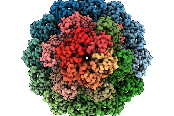 |
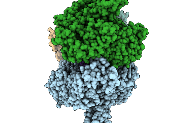
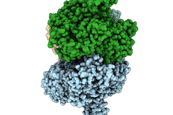
Structure Of Bacteriophage T4 Protal-Neck Protein Gp20-Gp13-Gp14-Hfq Assembled In Vitro In C6 Symmetry
Organism: Escherichia phage t4
Method: ELECTRON MICROSCOPY Resolution:2.91 Å Release Date: 2026-02-18 Classification: VIRAL PROTEIN |
Organism: Escherichia phage t4
Method: ELECTRON MICROSCOPY
Release Date: 2026-02-18
 |
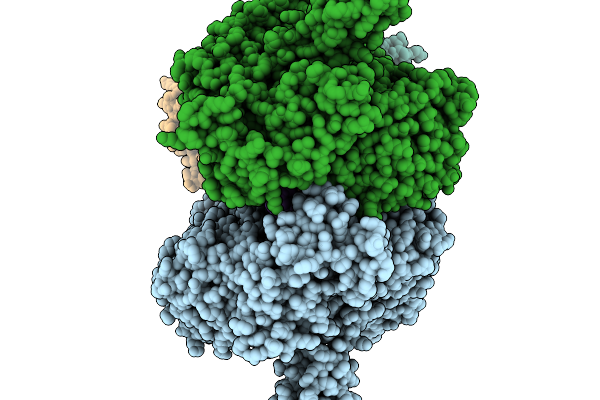
Structure Of Bacteriophage T4 Protal-Neck Mismatch Complex Gp20-Gp14-Gp13 Assembled In Vitro In C6 Symmetry
Organism: Escherichia phage t4
Method: ELECTRON MICROSCOPY Release Date: 2026-02-18 Classification: VIRAL PROTEIN |
Organism: Escherichia phage t4
Method: ELECTRON MICROSCOPY
Release Date: 2026-02-18
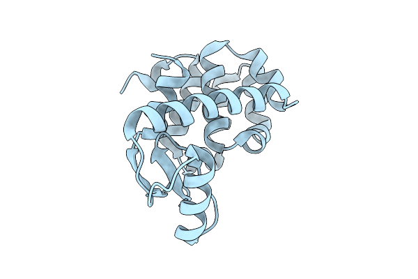 |
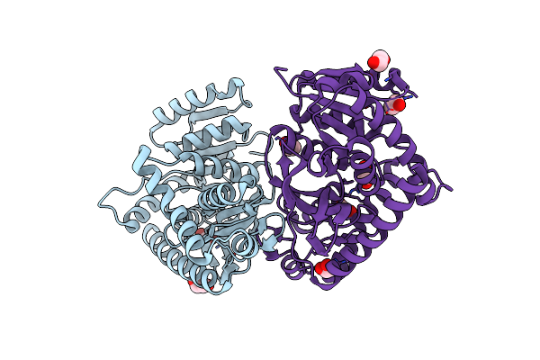
Spitrobot-2 Advances Time-Resolvedcryo-Trapping Crystallography To Under 25 Ms: T4 Lysozyme, Mutant L99A (Apo State)
Organism: Escherichia phage t4
Method: X-RAY DIFFRACTION Resolution:1.50 Å Release Date: 2025-12-03 Classification: HYDROLASE |
Organism: Escherichia phage t4
Method: X-RAY DIFFRACTION
Release Date: 2025-12-03
 |
Spitrobot-2 Advances Time-Resolvedcryo-Trapping Crystallography To Under 25 Ms: T4 Lysozyme, Mutant L99A Bound With Indole (1 S Soaking)
Organism: Escherichia phage t4
Method: X-RAY DIFFRACTION Resolution:1.90 Å Release Date: 2025-12-03 Classification: HYDROLASE Ligands: IND |
Organism: Escherichia phage t4
Method: X-RAY DIFFRACTION
Release Date: 2025-12-03
Ligands: IND
 |
Spitrobot-2 Advances Time-Resolvedcryo-Trapping Crystallography To Under 25 Ms: T4 Lysozyme, Mutant L99A Bound With Indole (10 S Soaking)
Organism: Escherichia phage t4
Method: X-RAY DIFFRACTION Resolution:1.49 Å Release Date: 2025-12-03 Classification: HYDROLASE Ligands: IND, NA |
Organism: Escherichia phage t4
Method: X-RAY DIFFRACTION
Release Date: 2025-12-03
Ligands: IND, NA
 |
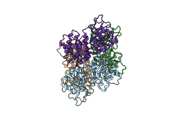
T4 Bacteriophage Replicative Polymerase Captured In Polymerase Exchange State 1
Organism: Escherichia phage t4, Synthetic construct
Method: ELECTRON MICROSCOPY Release Date: 2025-11-19 Classification: replication/DNA |
Organism: Escherichia phage t4, Synthetic construct
Method: ELECTRON MICROSCOPY
Release Date: 2025-11-19
 |
T4 Bacteriophage Replicative Polymerase Captured In Polymerase Exchange State 2
Organism: Escherichia phage t4, Synthetic construct
Method: ELECTRON MICROSCOPY Release Date: 2025-11-19 Classification: replication/DNA |
Organism: Escherichia phage t4, Synthetic construct
Method: ELECTRON MICROSCOPY
Release Date: 2025-11-19
 |
T4 Bacteriophage Replicative Holoenzyme Containing Triple Mutations D75R, Q430E, And K432E In The Exonuclease-Deficient Polymerase
Organism: Escherichia phage t4, Synthetic construct
Method: ELECTRON MICROSCOPY Release Date: 2025-11-19 Classification: Transferase/DNA |
Organism: Escherichia phage t4, Synthetic construct
Method: ELECTRON MICROSCOPY
Release Date: 2025-11-19
 |
T4 Bacteriophage Replicative Polymerase Captured In Polymerase Exchange State 3
Organism: Escherichia phage t4, Synthetic construct
Method: ELECTRON MICROSCOPY Release Date: 2025-11-19 Classification: Replication/DNA Ligands: CA, D3T |
Organism: Escherichia phage t4, Synthetic construct
Method: ELECTRON MICROSCOPY
Release Date: 2025-11-19
Ligands: CA, D3T
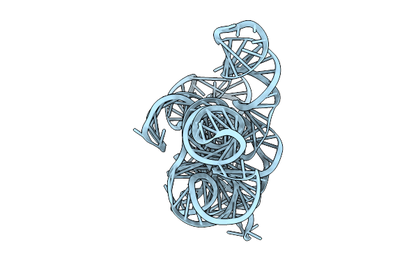 |
Cryo-Em Structure Of Linear Intron Of Thymidylate Synthase (Td) Gene Of Bacteriophage T4
Organism: Escherichia phage t4
Method: ELECTRON MICROSCOPY Release Date: 2025-11-12 Classification: RNA Ligands: MG |
Organism: Escherichia phage t4
Method: ELECTRON MICROSCOPY
Release Date: 2025-11-12
Ligands: MG
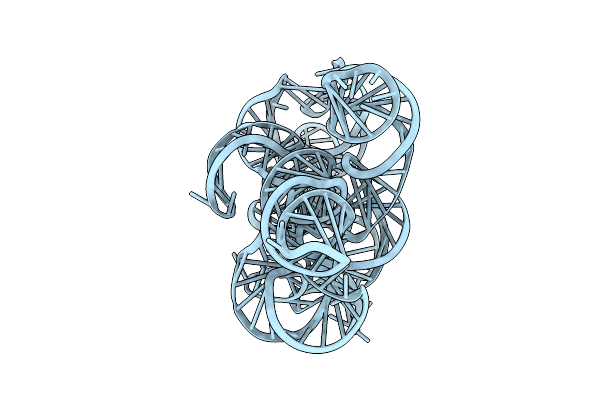 |
Cryo-Em Structure Of Circular Intron Of Thymidylate Synthase (Td) Gene Of Bacteriophage T4
Organism: Escherichia phage t4
Method: ELECTRON MICROSCOPY Release Date: 2025-11-12 Classification: RNA Ligands: MG |
Organism: Escherichia phage t4
Method: ELECTRON MICROSCOPY
Release Date: 2025-11-12
Ligands: MG
 |
Asgard Archaeal Hhob Nucleosome In The Closed Conformation
Organism: Candidatus heimdallarchaeota archaeon lc_3, Synthetic construct
Method: ELECTRON MICROSCOPY Resolution:3.50 Å Release Date: 2025-10-29 Classification: DNA BINDING PROTEIN |
Organism: Candidatus heimdallarchaeota archaeon lc_3, Synthetic construct
Method: ELECTRON MICROSCOPY
Release Date: 2025-10-29
 |
Asgard Hhob Hypernucleosome In The Closed State
Organism: Candidatus heimdallarchaeota archaeon lc_3, Synthetic construct
Method: ELECTRON MICROSCOPY Release Date: 2025-10-29 Classification: DNA BINDING PROTEIN |
Organism: Candidatus heimdallarchaeota archaeon lc_3, Synthetic construct
Method: ELECTRON MICROSCOPY
Release Date: 2025-10-29
 |
Asgard Hhob Nucleosome In The Open State
Organism: Candidatus heimdallarchaeota archaeon lc_3, Synthetic construct
Method: ELECTRON MICROSCOPY Resolution:3.60 Å Release Date: 2025-10-29 Classification: DNA BINDING PROTEIN |
Organism: Candidatus heimdallarchaeota archaeon lc_3, Synthetic construct
Method: ELECTRON MICROSCOPY
Release Date: 2025-10-29
 |
Glycosyltransferase C From The Limosilactobacillus Reuteri Accessory Secretion System. Apo Form.
Organism: Limosilactobacillus reuteri
Method: X-RAY DIFFRACTION Resolution:2.00 Å Release Date: 2025-04-23 Classification: TRANSFERASE Ligands: CA, GOL |
Organism: Limosilactobacillus reuteri
Method: X-RAY DIFFRACTION
Release Date: 2025-04-23
Ligands: CA, GOL
 |
Glycosyltransferase C From The Limosilactobacillus Reuteri Accessory Secretion System. Complex With Udp.
Organism: Limosilactobacillus reuteri
Method: X-RAY DIFFRACTION Resolution:2.60 Å Release Date: 2025-04-23 Classification: TRANSFERASE Ligands: UDP |
Organism: Limosilactobacillus reuteri
Method: X-RAY DIFFRACTION
Release Date: 2025-04-23
Ligands: UDP
 |
Crystal Structure L-Lactate Dehydrogenase From Lactobacillus Reuteri In Its Apoform
Organism: Limosilactobacillus reuteri
Method: X-RAY DIFFRACTION Resolution:2.15 Å Release Date: 2025-04-02 Classification: OXIDOREDUCTASE Ligands: EDO |
Organism: Limosilactobacillus reuteri
Method: X-RAY DIFFRACTION
Release Date: 2025-04-02
Ligands: EDO