

Search Count: 30
 |
Mbp-Mcl1 In Complex With Ligand 11
Organism: Serratia sp. fs14, Homo sapiens
Method: X-RAY DIFFRACTION Resolution:1.40 Å Release Date: 2023-05-17 Classification: APOPTOSIS Ligands: FMT, YLT |
Organism: Serratia sp. fs14, Homo sapiens
Method: X-RAY DIFFRACTION
Release Date: 2023-05-17
Ligands: FMT, YLT
 |
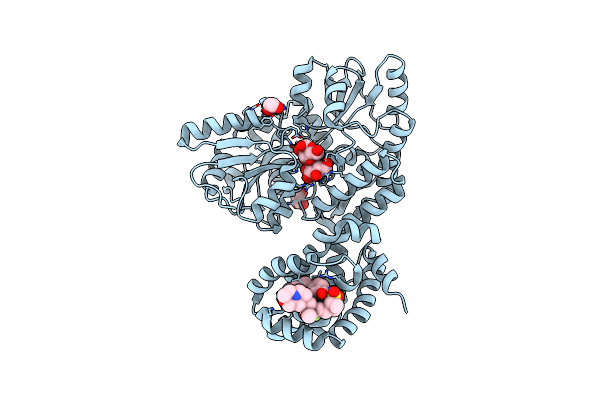
Mbp-Mcl1 In Complex With Ligand 12
Organism: Serratia sp. fs14, Homo sapiens
Method: X-RAY DIFFRACTION Resolution:1.83 Å Release Date: 2023-05-17 Classification: APOPTOSIS Ligands: FMT, EDO, YLK |
Organism: Serratia sp. fs14, Homo sapiens
Method: X-RAY DIFFRACTION
Release Date: 2023-05-17
Ligands: FMT, EDO, YLK
 |
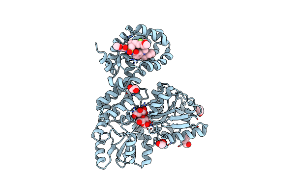
Mbp-Mcl1 In Complex With Ligand 21
Organism: Serratia sp. fs14, Homo sapiens
Method: X-RAY DIFFRACTION Resolution:1.94 Å Release Date: 2023-05-17 Classification: APOPTOSIS Ligands: EDO, YKT |
Organism: Serratia sp. fs14, Homo sapiens
Method: X-RAY DIFFRACTION
Release Date: 2023-05-17
Ligands: EDO, YKT
 |
Mbp-Mcl1 In Complex With Ligand 28
Organism: Serratia sp. fs14, Homo sapiens
Method: X-RAY DIFFRACTION Resolution:1.78 Å Release Date: 2023-05-17 Classification: APOPTOSIS Ligands: EDO, PEG, YKX |
Organism: Serratia sp. fs14, Homo sapiens
Method: X-RAY DIFFRACTION
Release Date: 2023-05-17
Ligands: EDO, PEG, YKX
 |
Mbp-Mcl1 In Complex With Ligand 32
Organism: Serratia sp. fs14, Homo sapiens
Method: X-RAY DIFFRACTION Resolution:1.46 Å Release Date: 2023-05-17 Classification: APOPTOSIS Ligands: EDO, YLF |
Organism: Serratia sp. fs14, Homo sapiens
Method: X-RAY DIFFRACTION
Release Date: 2023-05-17
Ligands: EDO, YLF
 |
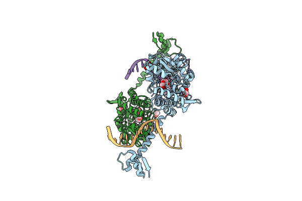
Mbp-Mcl1 In Complex With Ligand 34
Organism: Serratia sp. (strain fs14), Homo sapiens
Method: X-RAY DIFFRACTION Resolution:1.70 Å Release Date: 2023-05-17 Classification: APOPTOSIS Ligands: CL, EDO, PEG, YKL |
Organism: Serratia sp. (strain fs14), Homo sapiens
Method: X-RAY DIFFRACTION
Release Date: 2023-05-17
Ligands: CL, EDO, PEG, YKL
 |
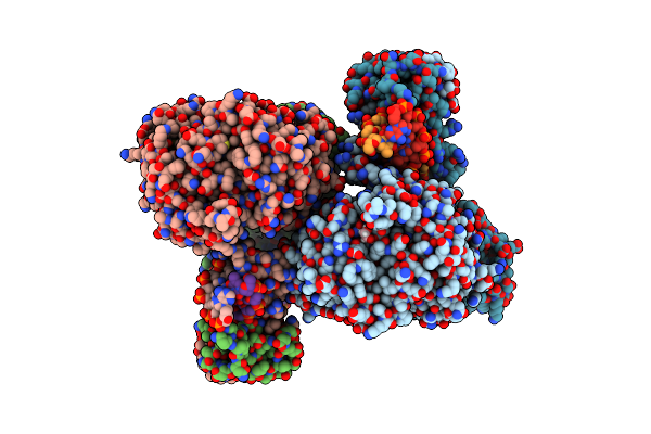
Crystal Structure Of Mbp-Fused Bil1/Bzr1 (21-90) In Complex With Double-Stranded Dna Contaning Atcacgtgat
Organism: Serratia sp. (strain fs14), Arabidopsis thaliana, Synthetic construct
Method: X-RAY DIFFRACTION Resolution:2.42 Å Release Date: 2022-12-07 Classification: TRANSCRIPTION Ligands: EDO |
Organism: Serratia sp. (strain fs14), Arabidopsis thaliana, Synthetic construct
Method: X-RAY DIFFRACTION
Release Date: 2022-12-07
Ligands: EDO
 |
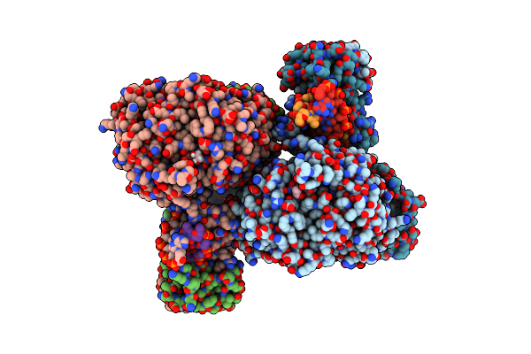
Crystal Structure Of Mbp-Fused Bil1/Bzr1 (21-90) In Complex With Double-Stranded Dna Contaning Cacacgtgtg
Organism: Serratia sp. (strain fs14), Arabidopsis thaliana, Synthetic construct
Method: X-RAY DIFFRACTION Resolution:1.94 Å Release Date: 2022-12-07 Classification: TRANSCRIPTION Ligands: EDO |
Organism: Serratia sp. (strain fs14), Arabidopsis thaliana, Synthetic construct
Method: X-RAY DIFFRACTION
Release Date: 2022-12-07
Ligands: EDO
 |
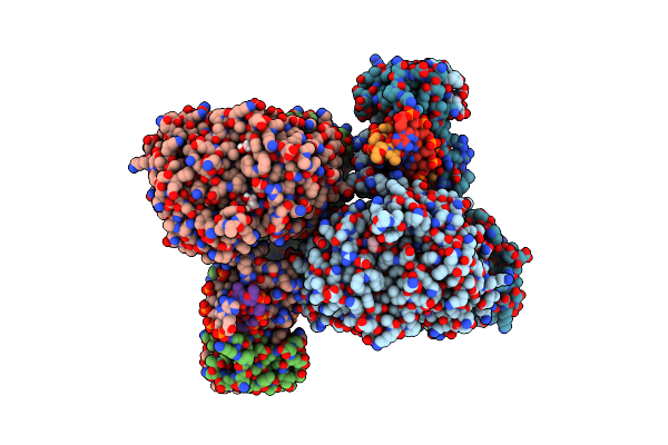
Crystal Structure Of Mbp-Fused Bil1/Bzr1 (21-90) In Complex With Double-Stranded Dna Contaning Tccacgtgga
Organism: Serratia sp. (strain fs14), Arabidopsis thaliana, Synthetic construct
Method: X-RAY DIFFRACTION Resolution:2.10 Å Release Date: 2022-12-07 Classification: TRANSCRIPTION Ligands: EDO |
Organism: Serratia sp. (strain fs14), Arabidopsis thaliana, Synthetic construct
Method: X-RAY DIFFRACTION
Release Date: 2022-12-07
Ligands: EDO
 |
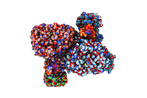
Crystal Structure Of Mbp-Fused Bil1/Bzr1 (21-90) In Complex With Double-Stranded Dna Contaning Ttcacgtgaa
Organism: Serratia sp. (strain fs14), Arabidopsis thaliana, Synthetic construct
Method: X-RAY DIFFRACTION Resolution:1.95 Å Release Date: 2022-12-07 Classification: TRANSCRIPTION Ligands: EDO |
Organism: Serratia sp. (strain fs14), Arabidopsis thaliana, Synthetic construct
Method: X-RAY DIFFRACTION
Release Date: 2022-12-07
Ligands: EDO
 |
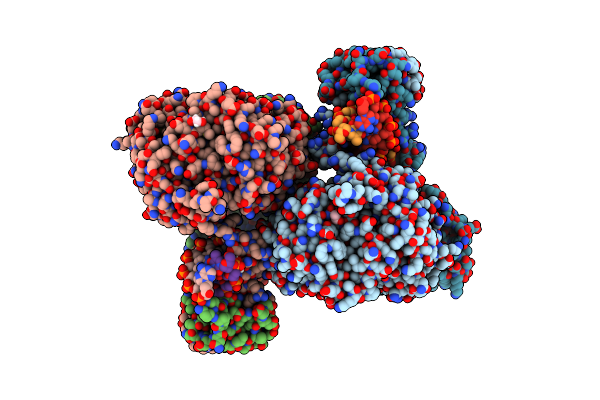
Crystal Structure Of Mbp-Fused Bil1/Bzr1 (21-90) In Complex With Double-Stranded Dna Contaning Cgcacgtgcg
Organism: Serratia sp. (strain fs14), Arabidopsis thaliana, Synthetic construct
Method: X-RAY DIFFRACTION Resolution:2.79 Å Release Date: 2022-12-07 Classification: TRANSCRIPTION Ligands: EDO |
Organism: Serratia sp. (strain fs14), Arabidopsis thaliana, Synthetic construct
Method: X-RAY DIFFRACTION
Release Date: 2022-12-07
Ligands: EDO
 |
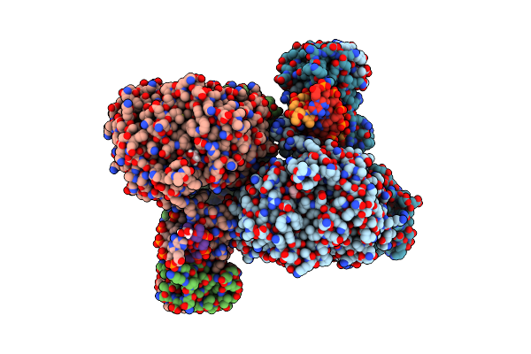
Crystal Structure Of Mbp-Fused Bil1/Bzr1 (21-90) In Complex With Double-Stranded Dna Contaning Gacacgtgtc
Organism: Serratia sp. (strain fs14), Arabidopsis thaliana, Synthetic construct
Method: X-RAY DIFFRACTION Resolution:2.11 Å Release Date: 2022-12-07 Classification: TRANSCRIPTION Ligands: EDO |
Organism: Serratia sp. (strain fs14), Arabidopsis thaliana, Synthetic construct
Method: X-RAY DIFFRACTION
Release Date: 2022-12-07
Ligands: EDO
 |
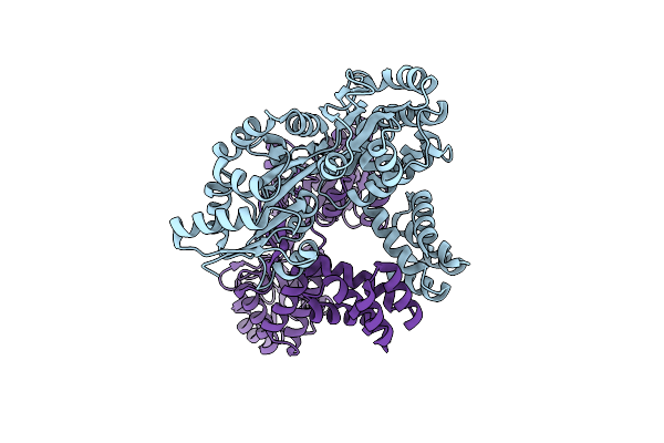
Crystal Structure Of Mbp-Fused Bil1/Bzr1 (21-90) In Complex With Double-Stranded Dna Contaning Gtcacgtgac
Organism: Serratia sp. (strain fs14), Arabidopsis thaliana, Synthetic construct
Method: X-RAY DIFFRACTION Resolution:2.04 Å Release Date: 2022-12-07 Classification: TRANSCRIPTION Ligands: EDO |
Organism: Serratia sp. (strain fs14), Arabidopsis thaliana, Synthetic construct
Method: X-RAY DIFFRACTION
Release Date: 2022-12-07
Ligands: EDO
 |
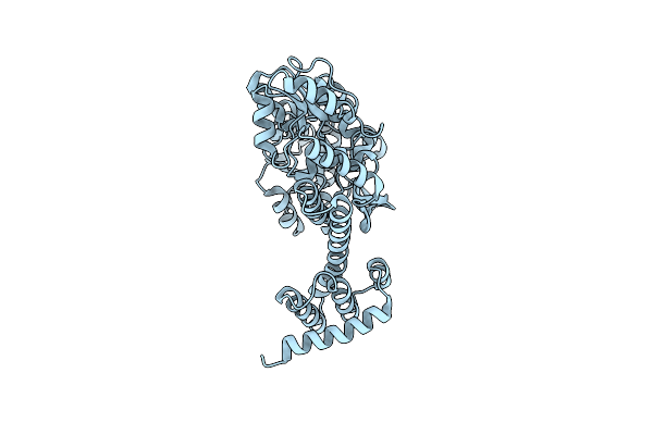
De Novo Design Protein D12 With Mbp Tag
Organism: Serratia sp. (strain fs14), Synthetic construct
Method: X-RAY DIFFRACTION Resolution:2.31 Å Release Date: 2021-12-22 Classification: UNKNOWN FUNCTION |
Organism: Serratia sp. (strain fs14), Synthetic construct
Method: X-RAY DIFFRACTION
Release Date: 2021-12-22
 |
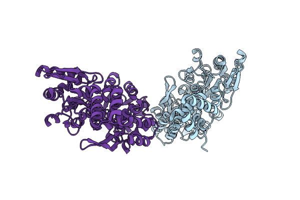
De Novo Design Protein D22 With Mbp Tag
Organism: Serratia sp. (strain fs14), Synthetic construct
Method: X-RAY DIFFRACTION Resolution:1.85 Å Release Date: 2021-12-22 Classification: UNKNOWN FUNCTION |
Organism: Serratia sp. (strain fs14), Synthetic construct
Method: X-RAY DIFFRACTION
Release Date: 2021-12-22
 |
De Novo Design Protein D53 With Mbp Tag
Organism: Serratia sp. (strain fs14), Synthetic construct
Method: X-RAY DIFFRACTION Resolution:2.85 Å Release Date: 2021-12-22 Classification: UNKNOWN FUNCTION |
Organism: Serratia sp. (strain fs14), Synthetic construct
Method: X-RAY DIFFRACTION
Release Date: 2021-12-22
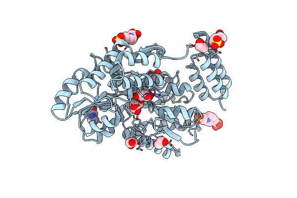 |
Structure Of The C-Terminal Domain Of The Menangle Virus Phosphoprotein (Residues 329 -388), Fused To Mbp. Space Group P212121
Organism: Serratia sp. (strain fs14), Menangle virus
Method: X-RAY DIFFRACTION Resolution:1.55 Å Release Date: 2021-10-06 Classification: VIRAL PROTEIN Ligands: EDO, PIN, PRO |
Organism: Serratia sp. (strain fs14), Menangle virus
Method: X-RAY DIFFRACTION
Release Date: 2021-10-06
Ligands: EDO, PIN, PRO
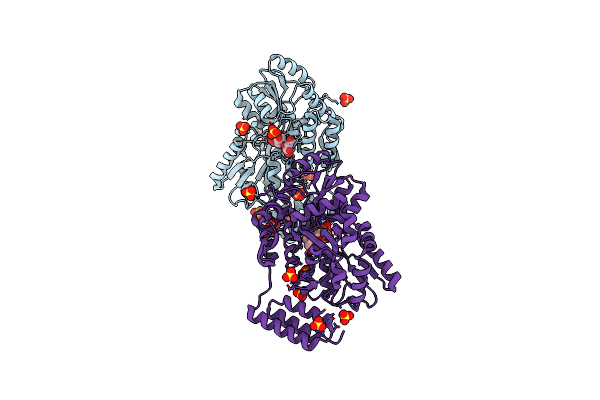 |
Structure Of The C-Terminal Domain Of The Menangle Virus Phosphoprotein (Residues 329 -388), Fused To Mbp. Space Group P21.
Organism: Serratia sp. (strain fs14), Menangle virus
Method: X-RAY DIFFRACTION Resolution:1.31 Å Release Date: 2021-09-15 Classification: VIRAL PROTEIN Ligands: SO4 |
Organism: Serratia sp. (strain fs14), Menangle virus
Method: X-RAY DIFFRACTION
Release Date: 2021-09-15
Ligands: SO4
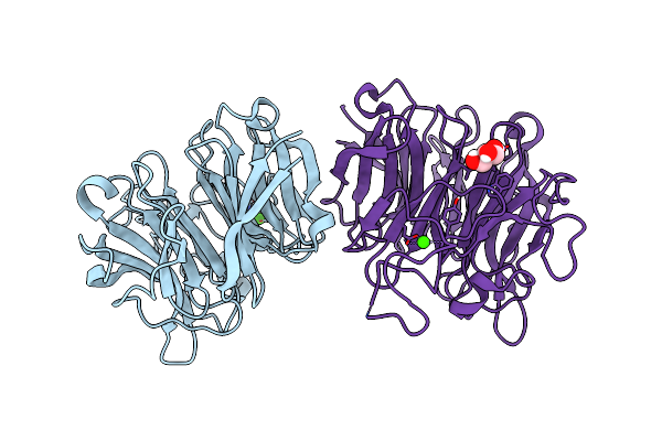 |
Glucose Dehydrogenase
Organism: Serratia sp. fs14
Method: X-RAY DIFFRACTION Resolution:1.94 Å Release Date: 2021-07-07 Classification: HYDROLASE Ligands: CA, GOL |
Organism: Serratia sp. fs14
Method: X-RAY DIFFRACTION
Release Date: 2021-07-07
Ligands: CA, GOL
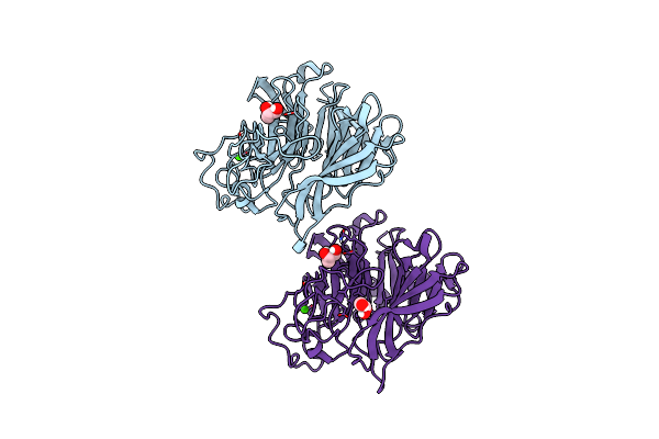 |
Crystal Structure Of Glucose Dehydrogenase
Organism: Serratia sp. fs14
Method: X-RAY DIFFRACTION Resolution:1.33 Å Release Date: 2021-06-23 Classification: HYDROLASE Ligands: GOL, CA |
Organism: Serratia sp. fs14
Method: X-RAY DIFFRACTION
Release Date: 2021-06-23
Ligands: GOL, CA