

Search Count: 32
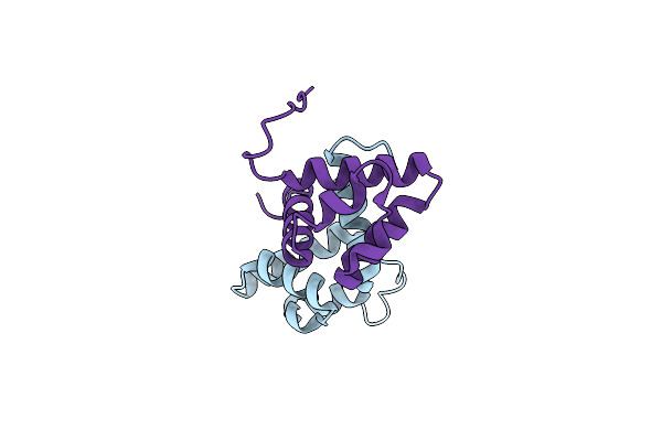 |
Ccps Mutant
Organism: Streptococcus suis
Method: X-RAY DIFFRACTION Resolution:1.50 Å Release Date: 2023-05-10 Classification: GENE REGULATION |
Organism: Streptococcus suis
Method: X-RAY DIFFRACTION
Release Date: 2023-05-10
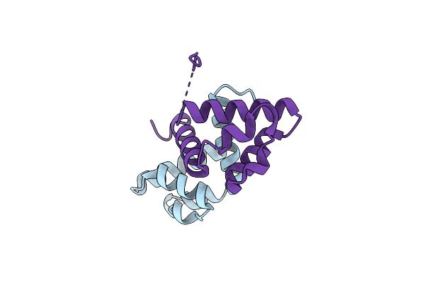 |
Ccps
Organism: Streptococcus suis
Method: X-RAY DIFFRACTION Resolution:1.60 Å Release Date: 2023-05-10 Classification: GENE REGULATION |
Organism: Streptococcus suis
Method: X-RAY DIFFRACTION
Release Date: 2023-05-10
 |
Structure Of Rdra From Streptococcus Suis Radar Defense System
Organism: Streptococcus suis
Method: ELECTRON MICROSCOPY Release Date: 2023-02-01 Classification: IMMUNE SYSTEM |
Organism: Streptococcus suis
Method: ELECTRON MICROSCOPY
Release Date: 2023-02-01
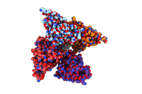 |
Streptococcus Suis Sadp Mutant - N285D
Organism: Streptococcus suis
Method: X-RAY DIFFRACTION Resolution:2.05 Å Release Date: 2020-08-19 Classification: SUGAR BINDING PROTEIN Ligands: GOL, NA |
Organism: Streptococcus suis
Method: X-RAY DIFFRACTION
Release Date: 2020-08-19
Ligands: GOL, NA
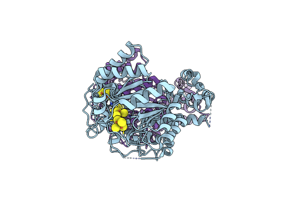 |
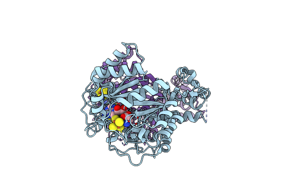
Crystal Structure Of Streptococcus Suis Suib
Organism: Streptococcus suis
Method: X-RAY DIFFRACTION Resolution:2.50 Å Release Date: 2017-08-30 Classification: METAL BINDING PROTEIN Ligands: SF4 |
Organism: Streptococcus suis
Method: X-RAY DIFFRACTION
Release Date: 2017-08-30
Ligands: SF4
 |
Crystal Structure Of Streptococcus Suis Suib Bound To S-Adenosylmethionine
Organism: Streptococcus suis
Method: X-RAY DIFFRACTION Resolution:2.49 Å Release Date: 2017-08-30 Classification: METAL BINDING PROTEIN Ligands: SF4, SAM |
Organism: Streptococcus suis
Method: X-RAY DIFFRACTION
Release Date: 2017-08-30
Ligands: SF4, SAM
 |
Crystal Structure Of Streptococcus Suis Suib Bound To Precursor Peptide Suia
Organism: Streptococcus suis
Method: X-RAY DIFFRACTION Resolution:2.10 Å Release Date: 2017-08-30 Classification: METAL BINDING PROTEIN Ligands: SF4, MET, SAM |
Organism: Streptococcus suis
Method: X-RAY DIFFRACTION
Release Date: 2017-08-30
Ligands: SF4, MET, SAM
 |
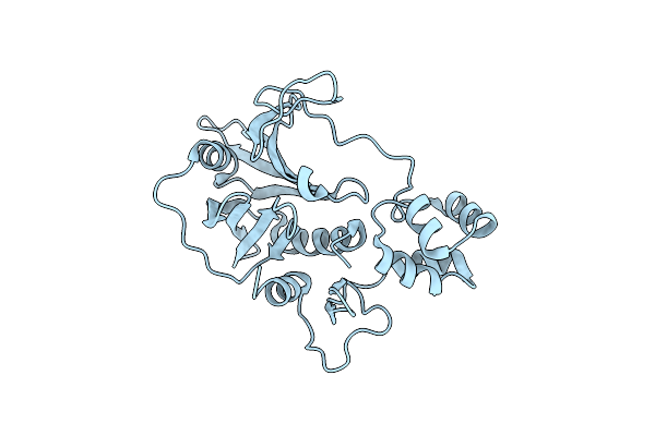
The Structure Of The C-Terminal Of The Fibronectin/Fibrinogen-Binding Protein From Streptococcus Suis (Fbps)
Organism: Streptococcus suis
Method: X-RAY DIFFRACTION Resolution:2.60 Å Release Date: 2016-11-16 Classification: CELL ADHESION |
Organism: Streptococcus suis
Method: X-RAY DIFFRACTION
Release Date: 2016-11-16
 |
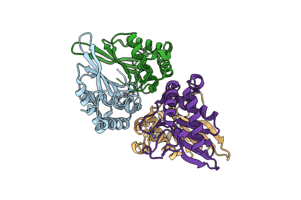
The Structure Of The N-Terminal Of The Fibronectin/Fibrinogen-Binding Protein From Streptococcus Suis (Fbps)
Organism: Streptococcus suis
Method: X-RAY DIFFRACTION Resolution:2.10 Å Release Date: 2016-11-16 Classification: CELL ADHESION |
Organism: Streptococcus suis
Method: X-RAY DIFFRACTION
Release Date: 2016-11-16
 |
S-Ribosylhomocysteinase From Streptococcus Suis
Organism: Streptococcus suis
Method: X-RAY DIFFRACTION Resolution:2.20 Å Release Date: 2015-06-24 Classification: LYASE Ligands: ZN |
Organism: Streptococcus suis
Method: X-RAY DIFFRACTION
Release Date: 2015-06-24
Ligands: ZN
 |
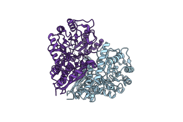
Crystal Structure Of A Tgase
Organism: Streptococcus suis
Method: X-RAY DIFFRACTION Resolution:2.10 Å Release Date: 2015-06-24 Classification: TRANSFERASE |
Organism: Streptococcus suis
Method: X-RAY DIFFRACTION
Release Date: 2015-06-24
 |
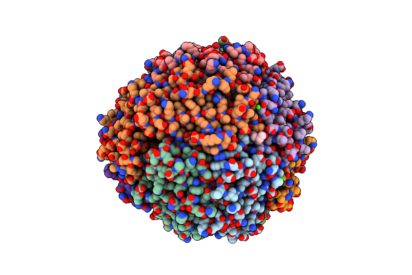
Structure Of The Enloase From Streptococcus Suis Serotype 2
Organism: Streptococcus suis
Method: X-RAY DIFFRACTION Resolution:2.40 Å Release Date: 2012-10-24 Classification: LYASE |
Organism: Streptococcus suis
Method: X-RAY DIFFRACTION
Release Date: 2012-10-24
 |
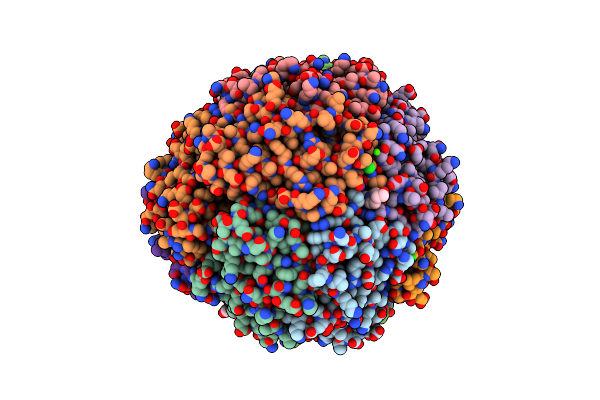
Crystal Structure Of Streptococcus Suis Dpr With Cobalt
Organism: Streptococcus suis
Method: X-RAY DIFFRACTION Resolution:2.30 Å Release Date: 2010-11-24 Classification: OXIDOREDUCTASE Ligands: CO, CL, CA, EPE |
Organism: Streptococcus suis
Method: X-RAY DIFFRACTION
Release Date: 2010-11-24
Ligands: CO, CL, CA, EPE
 |
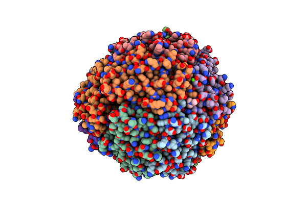
Crystal Structure Of Streptococcus Suis Dpr With Copper
Organism: Streptococcus suis
Method: X-RAY DIFFRACTION Resolution:2.10 Å Release Date: 2010-11-24 Classification: OXIDOREDUCTASE Ligands: CU, CL, EPE, CA |
Organism: Streptococcus suis
Method: X-RAY DIFFRACTION
Release Date: 2010-11-24
Ligands: CU, CL, EPE, CA
 |
Crystal Structure Of Streptococcus Suis Dpr With Nickel
Organism: Streptococcus suis
Method: X-RAY DIFFRACTION Resolution:2.10 Å Release Date: 2010-11-24 Classification: OXIDOREDUCTASE Ligands: NI, EPE, CA, CL |
Organism: Streptococcus suis
Method: X-RAY DIFFRACTION
Release Date: 2010-11-24
Ligands: NI, EPE, CA, CL
 |
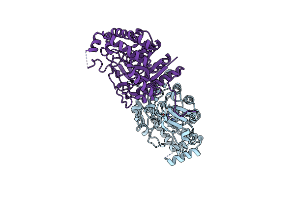
Crystal Structure Of Streptococcus Suis Dpr With Manganese
Organism: Streptococcus suis
Method: X-RAY DIFFRACTION Resolution:2.40 Å Release Date: 2010-11-24 Classification: OXIDOREDUCTASE Ligands: MN3, CL, EPE, CA |
Organism: Streptococcus suis
Method: X-RAY DIFFRACTION
Release Date: 2010-11-24
Ligands: MN3, CL, EPE, CA
 |
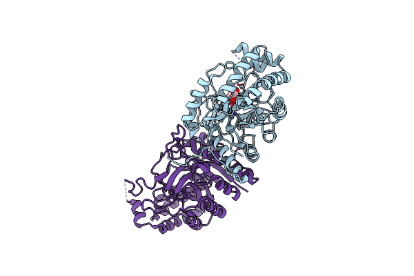
Crystal Structure Of Steptococcus Suis Mannonate Dehydratase With Metal Mn++
Organism: Streptococcus suis
Method: X-RAY DIFFRACTION Resolution:2.90 Å Release Date: 2009-09-01 Classification: LYASE Ligands: MN |
Organism: Streptococcus suis
Method: X-RAY DIFFRACTION
Release Date: 2009-09-01
Ligands: MN
 |
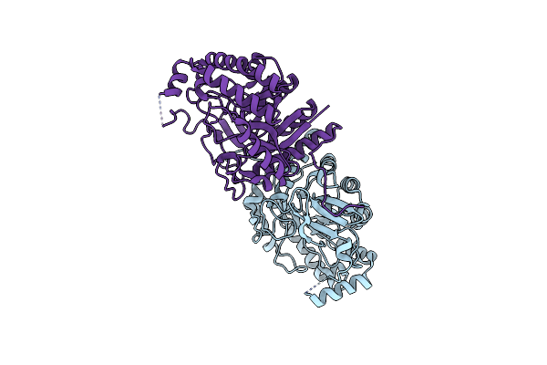
Crystal Structure Of Streptococcus Suis Mannonate Dehydratase Complexed With Substrate Analogue
Organism: Streptococcus suis
Method: X-RAY DIFFRACTION Resolution:2.50 Å Release Date: 2008-11-18 Classification: LYASE Ligands: DNO, MN |
Organism: Streptococcus suis
Method: X-RAY DIFFRACTION
Release Date: 2008-11-18
Ligands: DNO, MN
 |
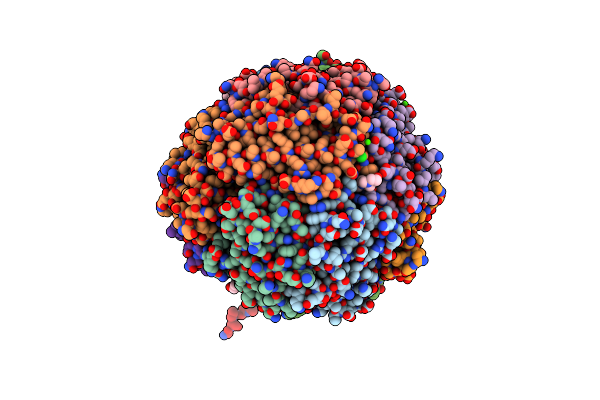
The Crystal Structure Of Mannonate Dehydratase From Streptococcus Suis Serotype2
Organism: Streptococcus suis
Method: X-RAY DIFFRACTION Resolution:2.90 Å Release Date: 2008-11-11 Classification: LYASE |
Organism: Streptococcus suis
Method: X-RAY DIFFRACTION
Release Date: 2008-11-11
 |
Terbium Binding In Streptococcus Suis Dpr Protein
Organism: Streptococcus suis
Method: X-RAY DIFFRACTION Resolution:2.10 Å Release Date: 2008-06-10 Classification: OXIDOREDUCTASE Ligands: CL, TB, CA, EPE |
Organism: Streptococcus suis
Method: X-RAY DIFFRACTION
Release Date: 2008-06-10
Ligands: CL, TB, CA, EPE