

Search Count: 12
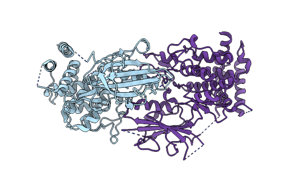 |
Structure Of Nucleotide-Free Pediculus Humanus (Ph) Pink1 Dimer
Organism: Pediculus humanus corporis
Method: ELECTRON MICROSCOPY Release Date: 2024-01-31 Classification: TRANSFERASE |
Organism: Pediculus humanus corporis
Method: ELECTRON MICROSCOPY
Release Date: 2024-01-31
 |
Structure Of Amp-Pnp-Bound Pediculus Humanus (Ph) Pink1 Dimer
Organism: Pediculus humanus corporis
Method: ELECTRON MICROSCOPY Release Date: 2024-01-31 Classification: TRANSFERASE Ligands: ANP, MG |
Organism: Pediculus humanus corporis
Method: ELECTRON MICROSCOPY
Release Date: 2024-01-31
Ligands: ANP, MG
 |
Structure Of Adp-Bound And Phosphorylated Pediculus Humanus (Ph) Pink1 Dimer
Organism: Pediculus humanus corporis
Method: ELECTRON MICROSCOPY Release Date: 2024-01-31 Classification: TRANSFERASE Ligands: ADP, MG |
Organism: Pediculus humanus corporis
Method: ELECTRON MICROSCOPY
Release Date: 2024-01-31
Ligands: ADP, MG
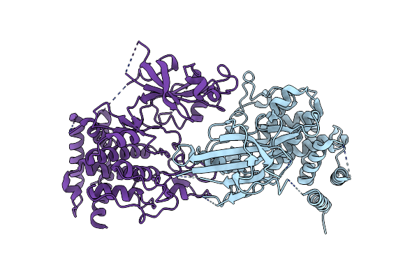 |
Structure Of Dimeric Phosphorylated Pediculus Humanus (Ph) Pink1 With Kinked Alpha-C Helix In Chain B
Organism: Pediculus humanus corporis
Method: ELECTRON MICROSCOPY Release Date: 2022-01-12 Classification: TRANSFERASE |
Organism: Pediculus humanus corporis
Method: ELECTRON MICROSCOPY
Release Date: 2022-01-12
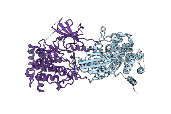 |
Structure Of Dimeric Phosphorylated Pediculus Humanus (Ph) Pink1 With Extended Alpha-C Helix In Chain B
Organism: Pediculus humanus corporis
Method: ELECTRON MICROSCOPY Release Date: 2022-01-12 Classification: TRANSFERASE |
Organism: Pediculus humanus corporis
Method: ELECTRON MICROSCOPY
Release Date: 2022-01-12
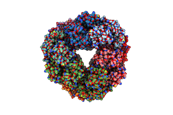 |
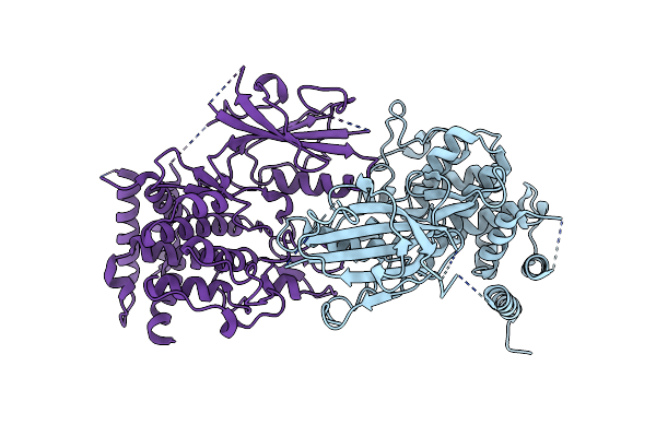
Structure Of Dodecameric Unphosphorylated Pediculus Humanus (Ph) Pink1 D357A Mutant
Organism: Pediculus humanus corporis
Method: ELECTRON MICROSCOPY Release Date: 2022-01-12 Classification: TRANSFERASE |
Organism: Pediculus humanus corporis
Method: ELECTRON MICROSCOPY
Release Date: 2022-01-12
 |
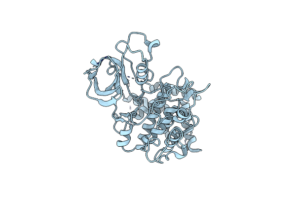
Structure Of Dimeric Unphosphorylated Pediculus Humanus (Ph) Pink1 D357A Mutant
Organism: Pediculus humanus corporis
Method: ELECTRON MICROSCOPY Release Date: 2022-01-12 Classification: TRANSFERASE |
Organism: Pediculus humanus corporis
Method: ELECTRON MICROSCOPY
Release Date: 2022-01-12
 |
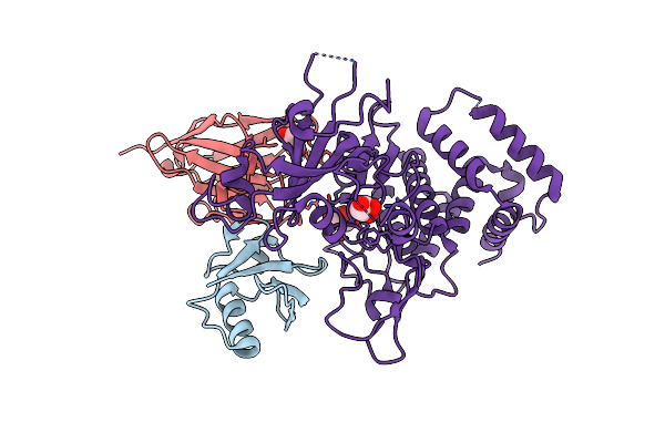
Structure Of Unphosphorylated Pediculus Humanus (Ph) Pink1 D334A Mutant
Organism: Pediculus humanus corporis
Method: X-RAY DIFFRACTION Resolution:3.53 Å Release Date: 2021-12-22 Classification: TRANSFERASE |
Organism: Pediculus humanus corporis
Method: X-RAY DIFFRACTION
Release Date: 2021-12-22
 |
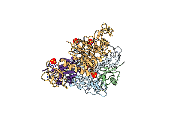
Structure Of Pink1 Bound To Ubiquitin
Organism: Homo sapiens, Lama glama, Pediculus humanus corporis
Method: X-RAY DIFFRACTION Resolution:3.10 Å Release Date: 2017-11-08 Classification: TRANSFERASE Ligands: GOL |
Organism: Homo sapiens, Lama glama, Pediculus humanus corporis
Method: X-RAY DIFFRACTION
Release Date: 2017-11-08
Ligands: GOL
 |
Structure Of Pediculus Humanus Parkin Bound To Phospho-Ubiquitin
Organism: Pediculus humanus subsp. corporis, Homo sapiens
Method: X-RAY DIFFRACTION Resolution:2.62 Å Release Date: 2015-07-22 Classification: SIGNALING PROTEIN Ligands: ZN, SO4 |
Organism: Pediculus humanus subsp. corporis, Homo sapiens
Method: X-RAY DIFFRACTION
Release Date: 2015-07-22
Ligands: ZN, SO4
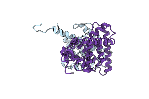 |
Crystal Structure Of The Ligand Binding Domains Of The Bovicola Ovis Ecdysone Receptor Ecr/Usp Heterodimer (Methylene Lactam Crystal)
Organism: Pediculus humanus subsp. corporis
Method: X-RAY DIFFRACTION Resolution:2.70 Å Release Date: 2014-07-30 Classification: TRANSCRIPTION |
Organism: Pediculus humanus subsp. corporis
Method: X-RAY DIFFRACTION
Release Date: 2014-07-30
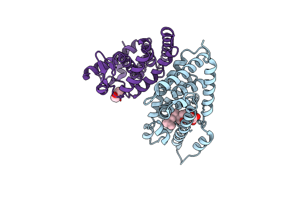 |
Crystal Structure Of The Ligand Binding Domains Of The Bovicola Ovis Ecdysone Receptor Ecr/Usp Heterodimer (Pona Crystal)
Organism: Pediculus humanus subsp. corporis
Method: X-RAY DIFFRACTION Resolution:2.70 Å Release Date: 2014-07-30 Classification: TRANSCRIPTION Ligands: P1A, NEQ |
Organism: Pediculus humanus subsp. corporis
Method: X-RAY DIFFRACTION
Release Date: 2014-07-30
Ligands: P1A, NEQ