IBIA: Indian Biological Images Archive
Image Data Submission Report
Generated on: 25 October 2026

| Project Accession: | IBIAP_1000000015 |
| Title: | DiverseCeph19: A Cephalometric Landmarks Annotation Dataset |
| Representative Image: | 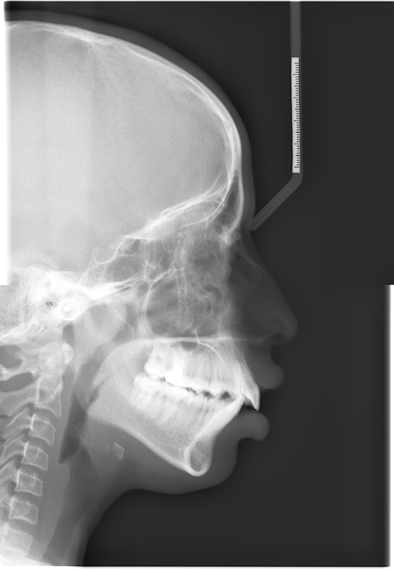 |
| Description: | Automation of cephalometric landmark annotation holds significant importance within the domain as a research area. This article presents a dataset consisting of 1692 cephalometric radiograph images, along with manually annotated data on 19 anatomical cephalometric landmarks. The data is available in individual text files corresponding to individual radiographic image files. Along with this, the latest annotation formats like COCO JSON and YOLO file formats for artificial intelligence (AI) model training are also available. Annotations are performed by experienced medical professionals with more than twenty years of experience and technical professionals with 6 years of research experience in the domain. The images are preprocessed and segregated based on resolution, image quality, dental structure, and other artifacts. The final image data is saved in BMP format with 0.127 mm/pixel resolution. The images refer to cephalometric analysis performed between 2011 and 2022 in patients treated at JSS Dental College and Hospital (JSS DCH), with patients age between 5 to 60 years. The demographic information of patients is not available with individual image data. Ethical clearance is obtained from the Institutional Ethical Committee of JSS DCH. Various levels of experimentation are carried out using the dataset, and the results demonstrate robust performance. Landmark annotation based on segregation type is one of the first types in this area of cephalometric landmark annotation. The availability of this dataset offers researchers a robust platform for investigating and conducting experiments using machine learning and deep learning techniques. |
| Publications: | https://doi.org/10.1016/j.compbiomed.2024.109318 |
| Associated Codes (URL only): | N/A |
| Funding agency: | N/A |
| Grant Number: | N/A |
| Ethics Statement: | Download |
| Any Other Information : | N/A |
| Additional File: | N/A |
| Acknowledgments: |
| Sr.No | First name | Last name | Organization | Designation | |
|---|---|---|---|---|---|
| 1 | Rashmi | S | rashmibe.nayak@gmail.com | JSS Science & Technology University, Mysuru | Research Scholar |
| 2 | Srinath | S | srinath@sjce.ac.in | JSS Science & Technology University, Mysuru | Principal Investigator |
| 3 | Prashanth | S | dr.prashanths@jssuni.edu.in | JSS Dental College & Hospital, Mysuru | Co-Principal Investigator |
| 4 | Seema | Deshmukh | dr.seemadeshmukh@jssuni.edu.in | JSS Dental College & Hospital, Mysuru | Co-Principal Investigator |
| 5 | Karthikeya | Patil | dr.karthikeyapatil@jssuni.edu.in | JSS Dental College & Hospital, Mysuru | Co-Principal Investigator |
| Study Accession: | XRS_1000000021 |
| Title: | Automation of Cephalometric Landmarks Annotation |
| Imaging Type: | Digital X-ray (XR) |
| Imaging Sub-type: | Diagnostic Radiology |
| Summary: | Each image collected from the hospital is first cropped to eliminate the patient-related information like name, sex, age, and date of image capture. Images are cropped to 2 distinct resolutions: 1341x1938 and 1257x1672 based on the lateral cephalometric region covered in the raw images and are saved in BMP format. Further, within individual resolution, images are segregated based on various factors, like 1. Teeth structure (supernumerary teeth, unerupted teeth) 2. Visibility of soft tissue profile. 3. Presence of orthodontic braces 4. Presence of earrings or a nose pin. Segregated image data is annotated by technical and medical professionals using ImageJ software (v1.53q). Identified landmarks are reviewed by experienced medical professionals, and finally, the landmark pixel locations are extracted into a text file. |
| Keywords: | Cephalometric radiographs; Anatomical Landmarks; Dataset; Machine Learning; Deep Learning |
| Additional / Any Other Information: | N/A |
| Release Date: | May 6, 2026 |
| Access Licence Type: | Open Access |
Table 1. The sample types registered under this study are as follows:
The total number of samples registered under this study is: 1692
| Sample Type ID | Organism | Taxon ID | Biological Entity | Laterality | Source Tissue | Source Cell/Cell-line | Cell Organelle |
|---|---|---|---|---|---|---|---|
| XRSMT_10000000049 | Homo sapiens | 9606 | Head | Right | NA | NA | NA |
Table 3. The experiment types registered under this study are as follows:
| Experiment Type ID | Instrument Name | Instrument Type | Manufacturer | Model |
|---|---|---|---|---|
| XRET_10000000019 | X-ray Machine | Planmeca Promax- Scara 3 | N/A | N/A |
| Experimental Design Summary (XRET_10000000019) |
|---|
| The images were derived from a cohort of 1557 subjects, consisting of 763 females and 794 males, spanning an age range from 5 to 60 years. The lateral cephalometric scans conducted over the period from 2011 to 2023 are considered for work. Utilizing the Planmeca Promax- Scara 3 machine with Romexis software, the images were originally saved in JPEG format, exhibiting varying image resolutions, with a pixel resolution of 0.127 mm/pixel. Patient names were used for image identification. To ensure anonymity and streamline the dataset for research purposes, the images underwent initial cropping to remove patient information located at the bottom of the cephalometric image which contained the subject’s details, age, and date of capture of the radiograph. Subsequently, the images are renamed with consecutive numerical identifiers. Following these preprocessing steps, the images are filtered based on different resolutions. Two major resolutions, 1341x1938 and 1257x1672, were selected to accommodate the diverse research requirements. These images were then converted and stored in BMP format. |
| Acquired Images Annotation Description (XRET_10000000019) |
|---|
| Three experienced professionals, each possessing over 20 years of expertise in the domain, manually annotated every image. Notably, the radiographs span the years 2012–2023, and the patients’ age range within the dataset extends from 7 to 50 years. |