IBIA: Indian Biological Images Archive
Image Data Submission Report
Generated on: 24 October 2026

| Project Accession: | IBIAP_1000000020 |
| Title: | Retinal Fundus Image Dataset for Multi Disease Biomarker Analysis |
| Representative Image: | 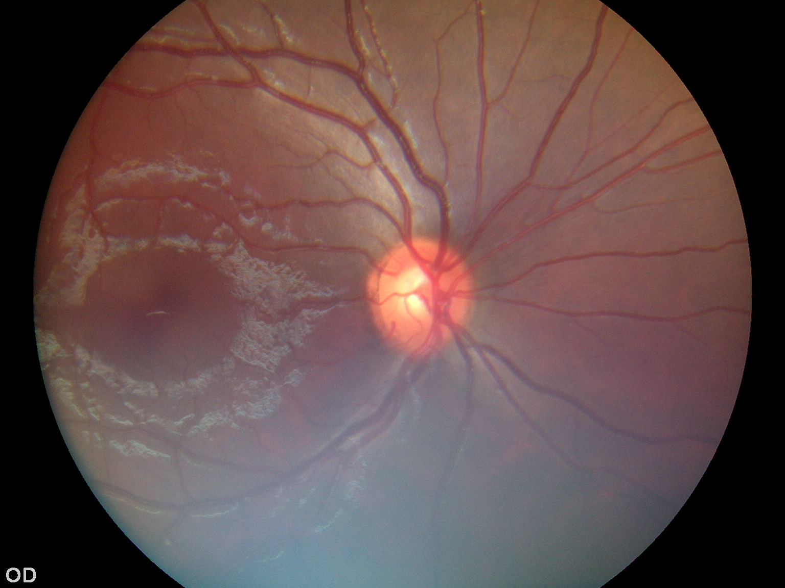 |
| Description: | This dataset presents the initial release of 500 non-mydriatic color fundus photographs from a larger ongoing collection aimed at investigating retinal biomarkers for systemic diseases, with a primary focus on cardiovascular disease and stroke. The images were collected at the Department of Ophthalmology, Amrita Institute of Medical Sciences, Kochi, India, between January 2024 and the present. All photographs were acquired using a handheld Zeiss Visuscout 100 fundus camera under standard, non-dilated clinical imaging conditions. Each participant underwent bilateral imaging, capturing both macula centered and optic disc centered views in accordance with clinical protocols. All images have been fully anonymized to remove any personally identifiable information. The images are stored in JPG format, with an average file size of 1.03 MB per image and a standardized resolution suitable for computational analysis. To support quantitative and automated research, retinal vessel segmentation and the identification of disease related patterns have been performed using advanced deep learning architectures. The current release serves as a representative subset of the full dataset, which will comprise over 10,000 images in future phases. Associated clinical metadata, including basic demographic details, relevant medical history, and laboratory results, are available to enhance the dataset’s research value. Given the clinical sensitivity of the data, access is restricted and will be provided upon request to qualified researchers for non-commercial, academic, or clinically relevant purposes. This dataset is intended for reuse in machine learning, image analysis, and algorithm development, with the goal of advancing understanding of the links between retinal health and systemic diseases in diverse populations. |
| Publications: | |
| Associated Codes (URL only): | N/A |
| Funding agency: | Indian Council of Medical Research, V. Ramalingaswami Bhawan, P.O. Box No. 4911, Ansari Nagar, New Delhi - 110029, India |
| Grant Number: | IIRP-2023-3943 |
| Ethics Statement: | Download |
| Any Other Information : | N/A |
| Additional File: | N/A |
| Acknowledgments: |
| Sr.No | First name | Last name | Organization | Designation | |
|---|---|---|---|---|---|
| 1 | Gopal S | Pillai | drgopalspillai@aims.amrita.edu | Amrita School of Medicine, Amrita Vishwa Vidyapeetham, Ponekkara P. O. Kochi, Kerala, India – 682041 | Principal Investigator |
| 2 | Merin | Dickson | merindm@aims.amrita.edu | Amrita School of Medicine, Amrita Vishwa Vidyapeetham, Ponekkara P. O. Kochi, Kerala, India – 682041 | Co-Investigator |
| 3 | Nagesh | Subbanna | nageshks@am.amrita.edu | Amrita Center for Wireless Networks and Applications (AWNA), Amrita Vishwa Vidyapeetham, Amritapuri Campus, Clappana P.O., Kollam – 690525, Kerala, India | Co-Principal Investigator |
| 4 | Aiswarya | A | aiswaryaasokan@aims.amrita.edu | Amrita School of Medicine, Amrita Vishwa Vidyapeetham, Ponekkara P. O. Kochi, Kerala, India – 682041 | Research Scholar |
| Study Accession: | OPTHS_1000000049 |
| Title: | RET-Amrita: Retinal Fundus Image Dataset |
| Imaging Type: | Ophthalmic Imaging (OPTH) |
| Imaging Sub-type: | None |
| Summary: | This dataset marks the initial release of 500 non-mydriatic retinal fundus photographs, part of an ongoing study into retinal biomarkers for systemic diseases like cardiovascular disease and stroke. Collected from January 2024 at the Amrita Institute of Medical Sciences in Kochi, India, the images were captured using a handheld Zeiss Visuscout 100 camera under standard clinical conditions. The collection is fully anonymized, standardized in resolution, and stored in JPG format. The current upload includes both normal retinal images and those exhibiting pathological features. While the cohort consists of 500 images, comprehensive clinical metadata (demographics, medical history, and lab results) is available for 230 patients. To facilitate deep learning development, expert annotations are provided for a specific subset of the data rather than the entire collection. These ground-truth masks include Vessel delineations for veins (n = 100) and arteries (n = 100). Pathology masks for cotton wool spots/soft exudates, hard exudates, and microaneurysms (n = 60), as well as hemorrhages (n = 60), Fibrous proliferation (n = 10), PRP Spots (10). This release is a curated subset of a larger project intended to reach over 10,000 images. The remaining data and supplemental annotations can be obtained by contacting the corresponding author. Access is restricted to qualified researchers for non-commercial, academic, or clinical use. This resource aims to advance precision diagnostics and predictive modeling in ophthalmology and systemic disease research. |
| Keywords: | Retinal fundus photography; Non-mydriatic imaging; Retinal biomarkers; Clinical metadata |
| Additional / Any Other Information: | Download |
| Release Date: | June 24, 2026 |
| Access Licence Type: | Open Access |
Table 1. The sample types registered under this study are as follows:
The total number of samples registered under this study is: 500
| Sample Type ID | Organism | Taxon ID | Biological Entity | Laterality | Source Tissue | Source Cell/Cell-line | Cell Organelle |
|---|---|---|---|---|---|---|---|
| OPTHSMT_10000000071 | Homo sapiens | 9606 | Eye | Both | Retina | N/A | N/A |
Table 3. The experiment types registered under this study are as follows:
| Experiment Type ID | Instrument Name | Instrument Type | Manufacturer | Model |
|---|---|---|---|---|
| OPTHET_10000000038 | Non-mydriatic Fundus Camera | Ophthalmic diagnostic imaging device | Zeiss | Zeiss visuscout 100 |
| Experimental Design Summary (OPTHET_10000000038) |
|---|
| This study explores the automated analysis of retinal fundus images to identify clinically relevant features and pathological patterns. The dataset consists of 500 non-mydriatic fundus images, captured using a handheld fundus camera under standardized clinical conditions. For a subset of patients, associated clinical metadata is available. Expert-annotated segmentation masks are provided for several retinal structures (e.g., arteries and veins) and pathological findings (e.g., microaneurysms, exudates, hemorrhages), although annotations are not uniformly available across all images. Supervised learning methods are employed for both classification and segmentation tasks, contingent upon the availability of segmentation masks. To mitigate challenges arising from class imbalance and heterogeneous annotation coverage, tailored sampling strategies are incorporated during training. Additionally, metadata regarding mask availability is leveraged to dynamically select valid supervision targets. This methodological design enables a rigorous evaluation of preprocessing strategies and model learning behaviour, thereby advancing clinically relevant applications of retinal image analysis. |
| Acquired Images Annotation Description (OPTHET_10000000038) |
|---|
| The dataset consists of 500 colour retinal fundus images collected from clinical ophthalmology records, representing both normal and pathological cases. Images are acquired under standardized conditions and stored in JPEG format, following a structured naming convention (opth_<IMAGE_ID>_<RECORD_ID>_<EYE>_FUNDUS.JPG) that encodes image index, patient record linkage, and laterality (left/right eye). The dataset includes 230 patients and corresponding clinical records, with a many-to-one relationship between patients and images. Pixel-level annotations are provided as segmentation masks in PNG format and organised into class-specific directories. The dataset supports multi-label annotation, allowing multiple retinal features to coexist within a single image. Annotated classes include retinal arteries and veins, as well as pathological features such as microaneurysms, hard and soft exudates, dot/blot and flame-shaped hemorrhages, fibrous proliferation, and pan-retinal photocoagulation scars. Annotation coverage is partial and heterogeneous: only 280 images contain at least one mask, and mask distribution varies significantly across classes, with arteries and veins being the most frequently annotated (100 images each), while rarer conditions such as fibrous proliferation and PRP scars have limited representation. The excel file specifies the presence or absence of each annotation type per image, enabling selective supervision during model training. In addition, a central metadata file (Metadata.csv) provides patient-level clinical labels, including age, gender, and systemic comorbidities such as diabetes, hypertension, coronary artery disease, and stroke. All dataset components are linked through a consistent unique identifier (opth_<IMAGE_ID>_<RECORD_ID>), ensuring precise integration of images, annotations, and clinical metadata for supervised and multimodal learning tasks. |