IBIA: Indian Biological Images Archive
Image Data Submission Report
Generated on: 04 October 2026

| Project Accession: | IBIAP_1000000044 |
| Title: | Indian Mammography Database 2.0 (IMDB 2.0) |
| Representative Image: | 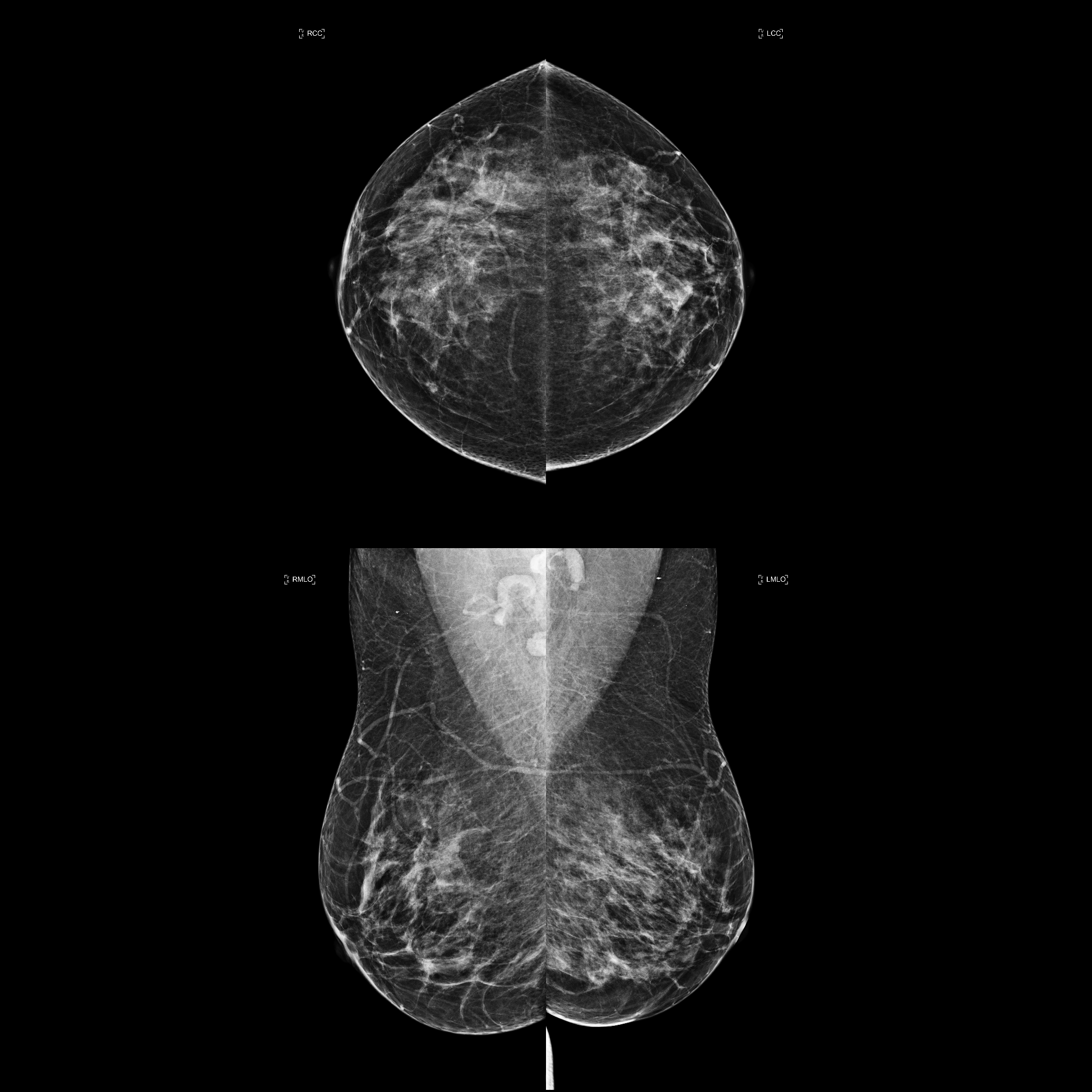 |
| Description: | This dataset consists of de-identified Full-Field Digital Mammography (FFDM) and Synthetic Mammography (SM) examinations acquired from women aged 18 years and older at a tertiary care oncology center between August and December 2025. The dataset includes BI-RADS assessments and breast density information extracted from radiology reports. Histopathology serves as the reference standard for suspicious lesions when available (BI-RADS 4/5), while follow-up imaging is available for probably benign cases (BI-RADS 3). Designed for artificial intelligence research, the dataset supports applications such as breast cancer detection, lesion classification, breast density assessment, and computer-aided diagnosis using deep learning and computer vision approaches. |
| Publications: | N/A |
| Associated Codes (URL only): | N/A |
| Funding agency: | N/A |
| Grant Number: | N/A |
| Ethics Statement: | Download |
| Any Other Information : | N/A |
| Additional File: | N/A |
| Acknowledgments: | The authors acknowledge the Department of Radiology, AIIMS New Delhi and Department of Oncoradiology, IRCH, AIIMS New Delhi, for technical and clinical support. The authors also acknowledge partial funding received by TANUH. |
| Sr.No | First name | Last name | Organization | Designation | |
|---|---|---|---|---|---|
| 1 | Varun | Holla | varunholla35@gmail.com | Dr. B.R.A.IRCH, All India Institute of Medical Sciences (AIIMS), New Delhi, India | Unspecified |
| 2 | Ashish | Rastogi | ashishrastogi.3150@gmail.com | Dr. B.R.A.IRCH, All India Institute of Medical Sciences (AIIMS), New Delhi, India | Unspecified |
| 3 | Omshivom | Nagpal | shivam.aiims.coe@gmail.com | Dr. B.R.A.IRCH, All India Institute of Medical Sciences (AIIMS), New Delhi, India | Unspecified |
| 4 | Kushagra | Chaturvedi | kushagrachaturvedi15@gmail.com | Dr. B.R.A.IRCH, All India Institute of Medical Sciences (AIIMS), New Delhi, India | Unspecified |
| 5 | Vipin | Thampi | thampi1990aiims@gmail.com | Dr. B.R.A.IRCH, All India Institute of Medical Sciences (AIIMS), New Delhi, India | Unspecified |
| 6 | Aditi | Madame | aditimadame19@gmail.com | Dr. B.R.A.IRCH, All India Institute of Medical Sciences (AIIMS), New Delhi, India | Unspecified |
| 7 | Hema | Malhotra | hemamalhotraaiims.2020@gmail.com | Dr. B.R.A.IRCH, All India Institute of Medical Sciences (AIIMS), New Delhi, India | Unspecified |
| 8 | Sathish | R | sathishappu314@gmail.com | Dr. B.R.A.IRCH, All India Institute of Medical Sciences (AIIMS), New Delhi, India | Unspecified |
| 9 | Krithika | Rangarajan | krithikarangarajan@aiims.edu | Dr. B.R.A.IRCH, All India Institute of Medical Sciences (AIIMS), New Delhi, India | Principal Investigator |
| Study Accession: | MAMOS_1000000050 |
| Title: | Indian Mammography Database 2.0 (IMDB 2.0) |
| Imaging Type: | Mammography (MAMO) |
| Imaging Sub-type: | Diagnostic Radiology |
| Summary: | This dataset consists of de-identified Full-Field Digital Mammography (FFDM) and Synthetic Mammography (SM) examinations acquired from women aged 18 years and older at a tertiary care oncology center between August and December 2025. The dataset includes BI-RADS assessments and breast density information extracted from radiology reports. Histopathology serves as the reference standard for suspicious lesions when available (BI-RADS 4/5), while follow-up imaging is available for probably benign cases (BI-RADS 3). Designed for artificial intelligence research, the dataset supports applications such as breast cancer detection, lesion classification, breast density assessment, and computer-aided diagnosis using deep learning and computer vision approaches. |
| Keywords: | Breast cancer; Mammography; Screening |
| Additional / Any Other Information: | N/A |
| Release Date: | June 18, 2026 |
| Access Licence Type: | Open Access |
Table 1. The sample types registered under this study are as follows:
The total number of samples registered under this study is: 9097
| Sample Type ID | Organism | Taxon ID | Biological Entity | Laterality | Source Tissue | Source Cell/Cell-line | Cell Organelle |
|---|---|---|---|---|---|---|---|
| MAMOSMT_10000000072 | Homo sapiens | 9606 | Breast | Both | N/A | N/A | N/A |
Table 3. The experiment types registered under this study are as follows:
| Experiment Type ID | Instrument Name | Instrument Type | Manufacturer | Model |
|---|---|---|---|---|
| MAMOET_10000000039 | Mammography Machine | Digital mammography | Hologic | Selenia |
| Experimental Design Summary (MAMOET_10000000039) |
|---|
| All mammographic examinations included in the present study were acquired using the Hologic Selenia Dimensions mammography system at our institution. The standard imaging protocol consisted of two-dimensional (2D) mammographic views for each breast, namely the cranio-caudal (CC) and medio-lateral oblique (MLO) projections, along with digital breast tomosynthesis (DBT) and synthesized mammography (SM) images. These image sets were obtained during a single breast compression using the system’s combination acquisition mode. When mammographic findings appeared suspicious or when breast density was expected to reduce the sensitivity of mammography, supplementary breast ultrasonography (US) was performed during the same setting. In such cases, the final radiological interpretation incorporated findings from both mammography and ultrasound examinations. |
| Acquired Images Annotation Description (MAMOET_10000000039) |
|---|
| Assessments were reported according to the fifth edition of the Breast Imaging Reporting and Data System (BI-RADS). A single BI-RADS category was assigned to each breast based on the most clinically significant abnormality identified on either mammography or ultrasound. Mammography reports underwent a double-reading process, with an initial interpretation by a radiology trainee followed by a review by an experienced breast imaging specialist. In instances of disagreement, the final assessment was determined by the specialist radiologist. For reference standard determination, a composite ground truth approach was adopted. Lesions categorized as BI-RADS 4 or BI-RADS 5 were verified through histopathological examination of the image-guided biopsy procedures. Histopathology outcomes were classified as either malignant (cancer) or benign (non-cancer). For BI-RADS 3 lesions, follow-up imaging studies were reviewed to assess lesion stability over time and the available follow-ups are uploaded. For BI-RADS 1 and BI-RADS 2 categories, available follow-up examinations and clinical records were reviewed whenever possible. Cases without evidence of malignancy during the observation period were categorized as non-cancer. The finalized ground-truth labels were linked to unique patient identifiers and stored in a structured dataset for subsequent analysis and model development. |