IBIA: Indian Biological Images Archive
Image Data Submission Report
Generated on: 24 October 2026

| Project Accession: | IBIAP_1000000024 |
| Title: | Developing Deep Learning Models for Quantitative Estimation of Reticulin Fibers in Bone Marrow Trephine Biopsy Specimens |
| Representative Image: | 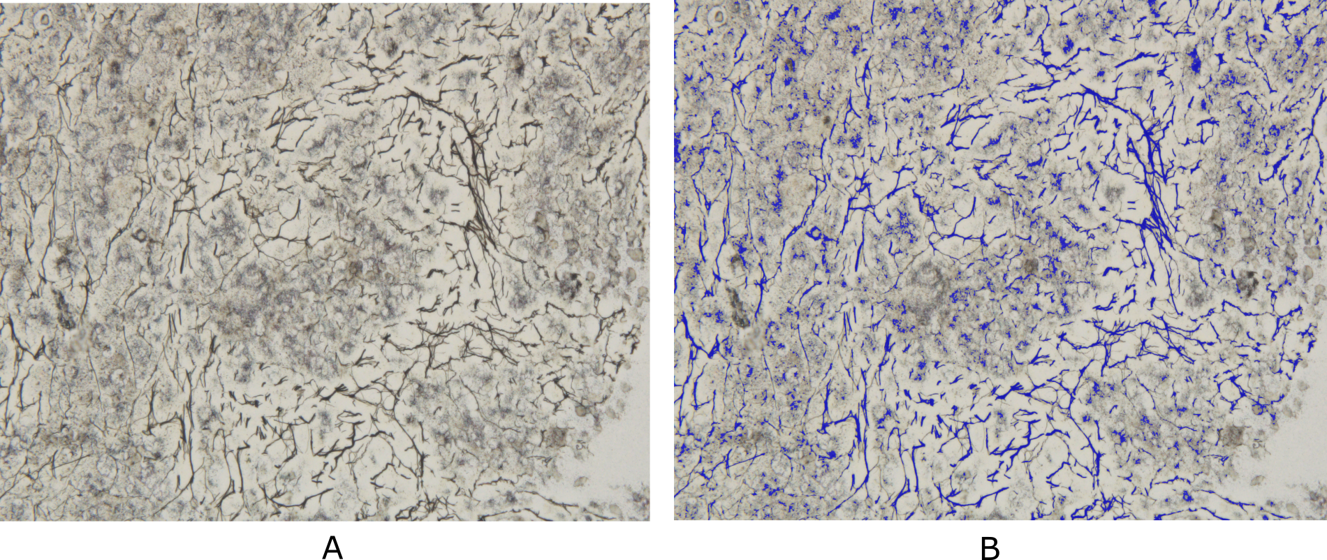 |
| Description: | Bone marrow reticulin fibrosis is associated with varied benign as well as malignant hematological conditions. The assessment of reticulin fibrosis is important in the diagnosis, prognostication and management of such disorders. The current methods for quantification of reticulin fibrosis are inefficient and prone to errors. Therefore, there is a need for automated tools for accurate and consistent quantification of reticulin. However, the lack of standardized datasets has hindered the development of such tools. In this study, we present a comprehensive dataset that comprises of 250 Bone Marrow Biopsy images for Reticulin (BoMBR) quantification. These images were meticulously annotated for semantic segmentation, with the focus on performing reticulin fiber quantification. This annotation was done by two trained hematopathologists who were aided by Deep Learning (DL) models and image processing techniques that generated a rough automated annotation for them to start with. This ensured precise delineation of the reticulin fibers alongside other cellular components such as bony trabeculae, fat, and cells. This is the first publicly available dataset in this domain with the aim to catalyze advancements the development of computational models for improved reticulin quantification. |
| Publications: | https://doi.org/10.59275/j.melba.2025-171e |
| Associated Codes (URL only): | N/A |
| Funding agency: | |
| Grant Number: | |
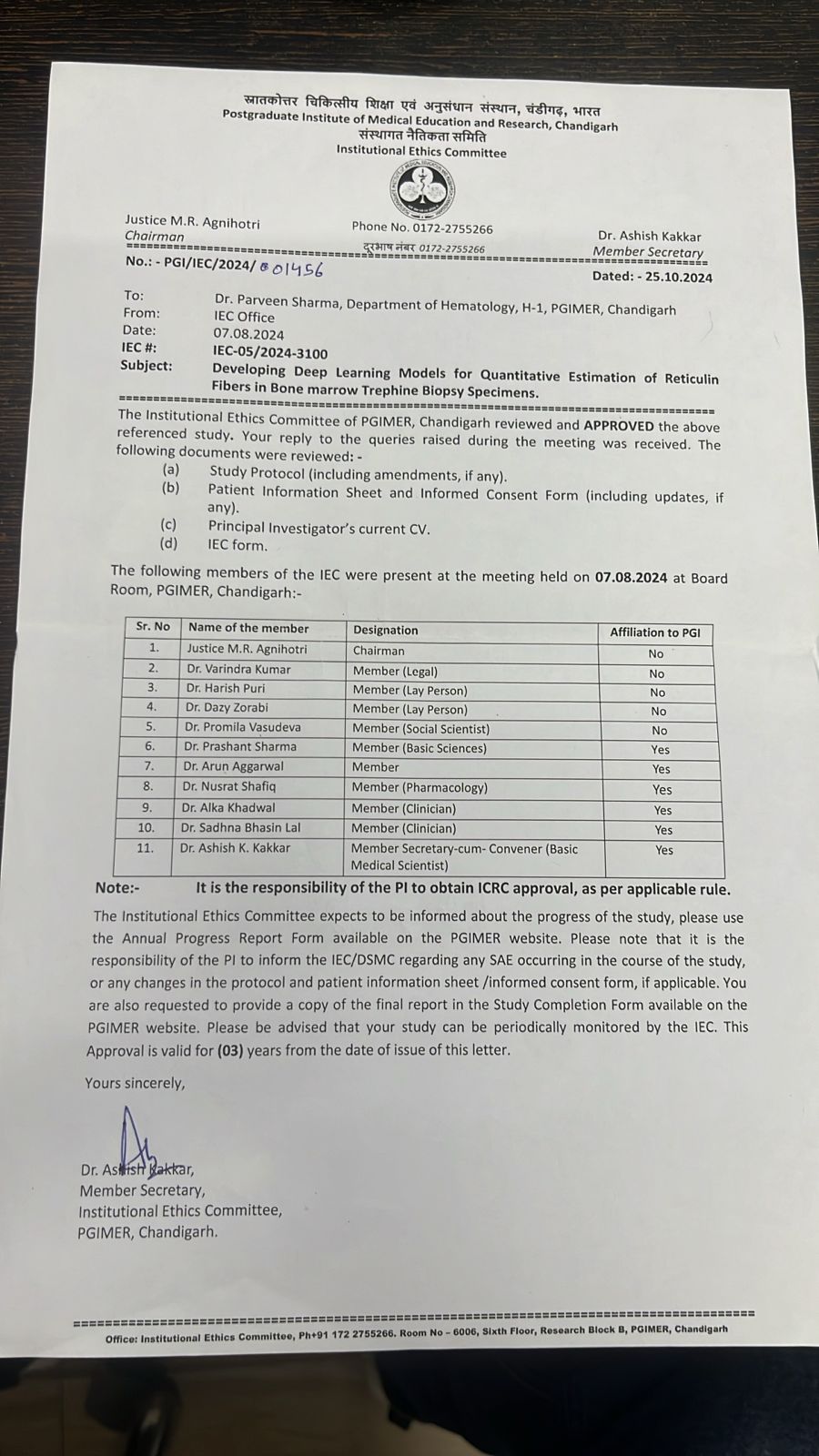
| Ethics Statement: | Download |
| Any Other Information : | The published article's link was updated on October 6, 2026. |
| Additional File: | N/A |
| Acknowledgments: |
| Sr.No | First name | Last name | Organization | Designation | |
|---|---|---|---|---|---|
| 1 | Sukrit | Gupta | sukrit.gupta@iitrpr.ac.in | IIT Ropar | Co-Principal Investigator |
| 2 | Praveen | Sharma | drsharmapgi@gmail.com | PGIMER, Chandigarh | Co-Principal Investigator |
| 3 | Satyender | Dharamdasani | sdharamdasani@gmail.com | PGIMER, Chandigarh | Co-Investigator |
| 4 | Panav | Raina | panavraina02@gmail.com | Panjab University | Co-Investigator |
| Study Accession: | HISTOS_1000000028 |
| Title: | BoMBR: Bone Marrow Biopsy Dataset for Segmentation of Reticulin Fibers |
| Imaging Type: | Histopathology (HISTO) |
| Imaging Sub-type: | Diagnostic Pathology |
| Summary: | We propose the BoMBR dataset containing 250 Bone Marrow Trephine (BMT) pixel-wise annotated images. Besides the pixel-wise annotation masks, we give information regarding the grade of Marrow Fibrosis, percentage cellular area covered by reticulin, and the average value of the elongation factor of the reticulin fibers in the image. The percentage of cellular area covered by reticulin indicates differences in the amount of area covered by reticulin fibers across different grades, while the elongation factor helps understand changes in the shape of reticulin fibers as the grade increases. This is by far the first such publicly available resource for annotated BMT images that focuses on reticulin fibrosis both globally and in the context of the Indian subcontinent. Our dataset aims to facilitate the effective and objective quantitative measurement of reticulin fibers, moving beyond the current qualitative, observer-dependent scoring systems. Beyond its primary application in the grading of myelofibrosis, the dataset may also serve as a benchmark for further studies. This includes its potential to assist hematopathologists in the classification of Myeloproliferative Neoplasms, assessment of disease progression, and quantitative monitoring of the effects of myelofibrosis reversal in patients, particularly in the context of clinical trials and novel targeted therapies. The annotated dataset of BMT biopsy images presented in this study provides a valuable resource for quantitative assessment of fibrosis severity in hematological disorders. By leveraging automated segmentation followed by expert annotation, we have created a comprehensive dataset covering a range of Marrow Fibrosis grades (MF-0 to MF-3). This dataset includes detailed annotations for reticulin fibers, bony trabeculae, fat regions, and cellular region, facilitating in-depth analysis of bone marrow pathology. |
| Keywords: | Medical Imaging; Reticulin Fibers; Marrow Fibrosis Grade Detection; Bone Marrow Trephine; Digital Pathology |
| Additional / Any Other Information: | N/A |
| Release Date: | Sept. 23, 2025 |
| Access Licence Type: | Open Access |
Table 1. The sample types registered under this study are as follows:
The total number of samples registered under this study is: 250
| Sample Type ID | Organism | Taxon ID | Biological Entity | Laterality | Source Tissue | Source Cell/Cell-line | Cell Organelle |
|---|---|---|---|---|---|---|---|
| HISTOSMT_10000000060 | Homo sapiens | 9606 | Bone Marrow | Not Applicable | Bone Marrow | N/A | N/A |
Table 3. The experiment types registered under this study are as follows:
| Experiment Type ID | Instrument Name | Instrument Type | Manufacturer | Model |
|---|---|---|---|---|
| HISTOET_10000000026 | Digital Slide Scanning System | Digital Slide Scanner | Motic | VM1 |
| Experimental Design Summary (HISTOET_10000000026) |
|---|
| Silver stained bone marrow trephine biopsy images. The BMT biopsy data was acquired using standardized procedures at the Postgraduate Institute of Medical Education and Research, Chandigarh (PGIMER). These procedures are as per medical standards as mentioned in Bancroft's Theory and Practice of Histological Techniques. After aspiration, samples underwent decalcification, fixation and paraffin embedding. Then biopsy specimens were prepared for microscopic examination by sectioning into 2-3 microns samples and staining with reticulin stain based on silver impregnation technique. High-resolution digital images of stained biopsy sections were captured using an Olympus BX53 microscope at a magnification of 400x and 1000x. A total of 19 patients diagnosed with MPN participated in the study. The time taken for data collection varied depending on various factors, including sample preparation and imaging. The time taken for sample preparation was approximately 120 minutes. For taking an image it takes approximately 10 seconds. The annotation of the images depends on the MF grade and ranges from 5 minutes to 20 minutes for MF-0 to MF-3. |
| Acquired Images Annotation Description (HISTOET_10000000026) |
|---|
| In our dataset, we ensured comprehensive representation across all myelofibrosis MF grades, encompassing a diverse range of pathological conditions. Specifically, we included 39 images of MF-0, 47 images of MF-1, 78 images of MF-2, and 37 images of MF-3. Hemorrhages by the leakage of blood from ruptured blood vessels into surrounding tissues or as bone marrow aspiration induced ones, are a common feature observed in bone marrow pathology. Our dataset initially comprised 202 images, but we discarded 1 image due to excessive hemorrhage, leaving us with a total of 201 images. This dataset features 55 images exhibiting hemorrhages, capturing its diverse manifestations within the bone marrow environment. By incorporating a wide range of MF grades and cellular samples, our dataset offers researchers a comprehensive resource for studying the complex interplay between fibrosis, cellular composition, and disease progression in MPN and other disease states. Besides this, we provided semantic segmentation for different types of regions in the BMT images, viz. Bony Trabecule, Fat, Cell, and Reticulin Fibers. The annotation process for the bone marrow biopsy images combined automated methods with manual verification to achieve accurate and reliable annotations. This hybrid approach leveraged the strengths of automated segmentation techniques while mitigating potential inaccuracies through expert intervention. The resulting annotated dataset, available in Pascal VOC format and annotated using CVAT, serves as a valuable resource for research and development in bone marrow evaluation and related fields. |