IBIA: Indian Biological Images Archive
Image Data Submission Report
Generated on: 26 October 2026

| Project Accession: | IBIAP_1000000023 |
| Title: | Developing a Deep Learning Framework for Clinical Use in Estimating Cellularity in Bone Marrow Biopsy Specimens |
| Representative Image: | 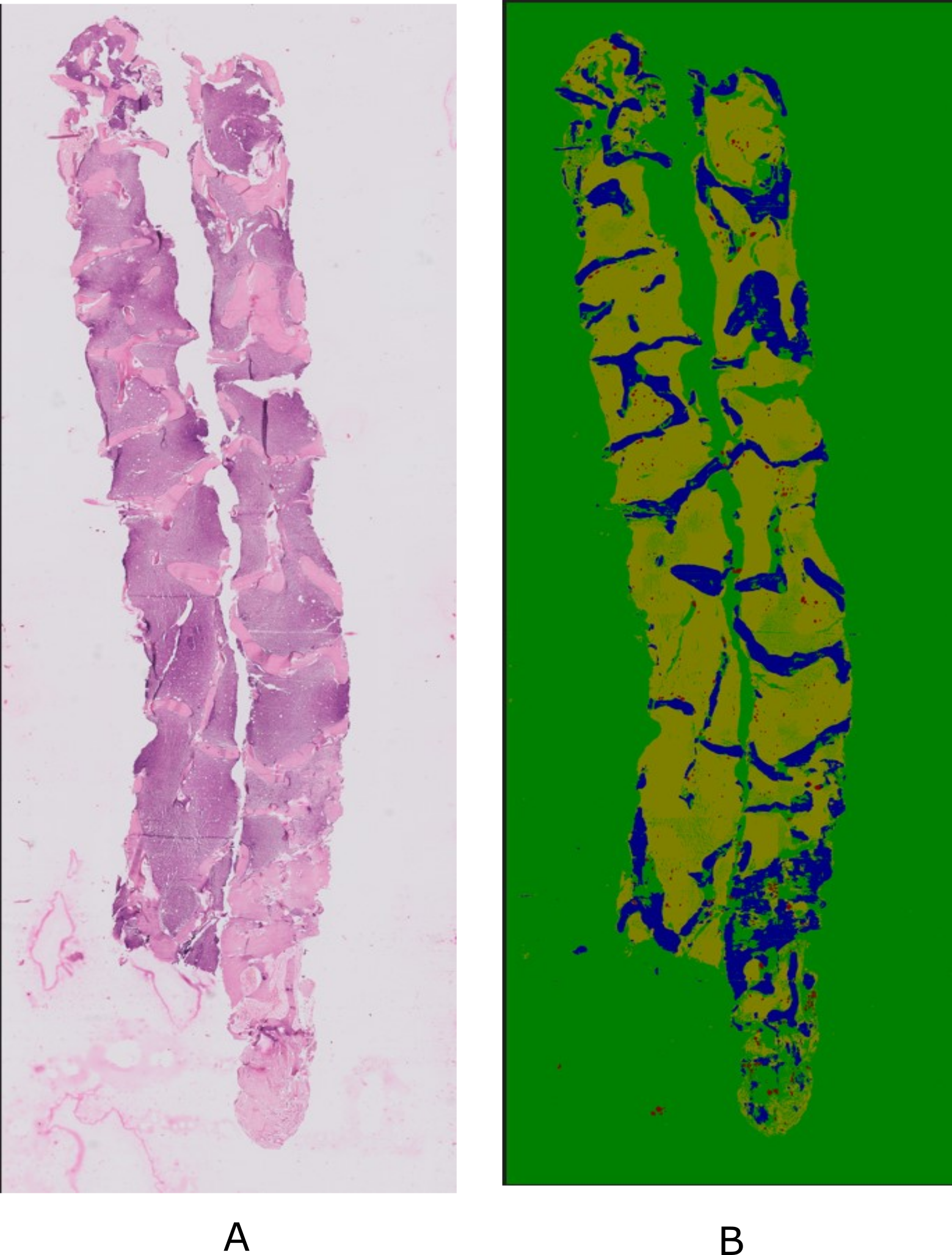 |
| Description: | Bone marrow examination has become increasingly important for the diagnosis and treatment of hematologic and other illnesses. The present methods for analyzing bone marrow biopsy samples involve subjective and inaccurate assessments by visual estimation by pathologists. Thus, there is a need to develop automated tools to assist in the analysis of bone marrow samples. However, there is a lack of publicly available standardized and high-quality datasets that can aid in the research and development of automated tools that can provide consistent and objective measurements. In this paper, we present a comprehensive Bone Marrow Biopsy (BaMBo) dataset consisting 229 semantic-segmented bone marrow biopsy images, specifically designed for the automated calculation of bone marrow cellularity. Our dataset comprises high-resolution, generalized images of bone marrow biopsies, each annotated with precise semantic segmentation of different haematological components. These components are divided into 4 classes: Bony trabeculae, adipocytes, cellular region and Background. The annotations were performed with the help of two experienced hematopathologists that were supported by state-of-the-art DL models and image processing techniques. |
| Publications: | https://doi.org/10.59275/j.melba.2025-dcae |
| Associated Codes (URL only): | N/A |
| Funding agency: | N/A |
| Grant Number: | N/A |
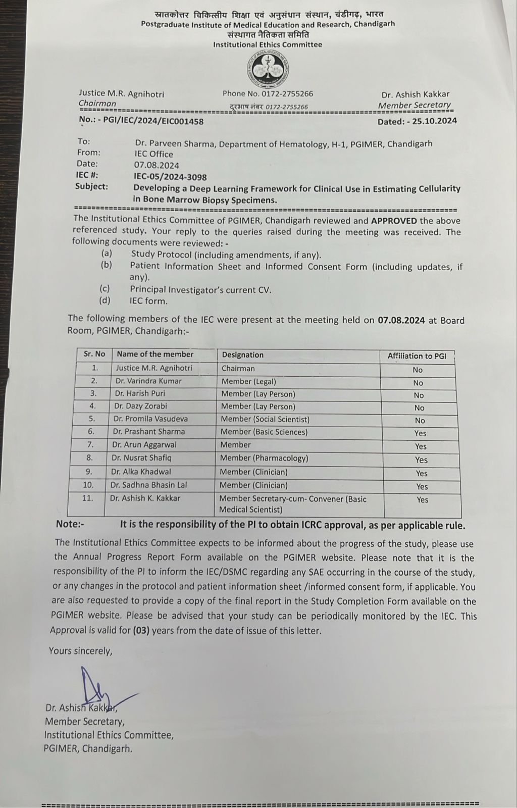
| Ethics Statement: | Download |
| Any Other Information : | The published article's link was updated on October 6, 2026. |
| Additional File: | N/A |
| Acknowledgments: | N/A |
| Sr.No | First name | Last name | Organization | Designation | |
|---|---|---|---|---|---|
| 1 | Praveen | Sharma | drsharmapgi@gmail.com | Postgraduate Institute of Medical Education and Research (PGIMER), Chandigarh, India | Co-Principal Investigator |
| 2 | Satyender | Dharamdasani | sdharamdasani@gmail.com | Postgraduate Institute of Medical Education and Research (PGIMER), Chandigarh, India | Co-Investigator |
| 3 | Sukrit | Gupta | sukrit.gupta@iitrpr.ac.in | Indian Institute of Technology (IIT), Ropar, Rupnagar, Punjab, India | Co-Principal Investigator |
| Study Accession: | HISTOS_1000000027 |
| Title: | BaMBo: An Annotated Bone Marrow Biopsy Dataset for Segmentation Task |
| Imaging Type: | Histopathology (HISTO) |
| Imaging Sub-type: | Diagnostic Pathology |
| Summary: | Data collection involved the acquisition of bone marrow trephine (BMT) biopsy specimens using standardized procedures at Postgraduate Institute of Medical Education and Research (PGIMER), Chandigarh. These procedures, as described later, are medical standards. The dataset comprises 229 biopsy images from a total of 38 patients, diagnosed with varied hematological states covering myeloproliferative neoplasms, leukemia, aplastic anemia, uninvolved staging marrows among others. Subjects were chosen at random from both the genders, contains a large age gap and varied medical history. All the patient related identifiers were removed from the dataset and the dataset was completely anonymized for patient confidentiality. |
| Keywords: | Medical imaging; Dataset; Bone marrow biopsy; Cellularity; Semi-automatic annotation |
| Additional / Any Other Information: | N/A |
| Release Date: | Oct. 16, 2025 |
| Access Licence Type: | Open Access |
Table 1. The sample types registered under this study are as follows:
The total number of samples registered under this study is: 229
| Sample Type ID | Organism | Taxon ID | Biological Entity | Laterality | Source Tissue | Source Cell/Cell-line | Cell Organelle |
|---|---|---|---|---|---|---|---|
| HISTOSMT_10000000059 | Homo sapiens | 9606 | Bone Marrow | Not Applicable | Bone Marrow | N/A | NA |
Table 3. The experiment types registered under this study are as follows:
| Experiment Type ID | Instrument Name | Instrument Type | Manufacturer | Model |
|---|---|---|---|---|
| HISTOET_10000000025 | Digital Slide Scanning System | Digital Slide Scanner | Motic | VM1 |
| Experimental Design Summary (HISTOET_10000000025) |
|---|
| Biopsies are taken from the posterior superior iliac spine. During the procedure, a larger needle was employed to remove a small core of the marrow tissue (biopsy) (Cloos et al., 2018; Rindy and Chambers, 2020). Biopsy samples then underwent Decalcification, Fixation and Paraffin embedding. They were then prepared for microscopic examination by sectioning into 2-3 microns samples and staining with Hematoxylin and Eosin (H&E). High-resolution digital images of stained biopsy sections were captured using an Olympus BX53 microscope at a magnification of 100x, 200x and 400x. The data collection was conducted over a period of three months and resulted in around 300 images. Out of the 300 bone marrow biopsy images obtained from PGIMER, images with poor clarity and repetitive images were excluded. After thorough inspection, 229 high quality images were retained. |
| Acquired Images Annotation Description (HISTOET_10000000025) |
|---|
| In order to perform the annotation of our biopsy specimens, we developed a preprocessing pipeline for first generating a rough annotation of the images. This pipeline made the annotation process faster and more efficient as the pathologist did not need to start from scratch. For this pipeline, we utilized a state-of-the-art segmentation model SAM (Kirillov et al., 2023) and the Computer Vision Annotation Tool (CVAT) platform for final annotation by the pathologist. SAM is a promptable segmentation system with zero-shot generalization to unfamiliar objects and images, without the need for additional training. The automated annotation pipeline gave us a rough annotation of each image. It is important to note that these annotations were not devoid of inaccuracies and that this was expected. These inaccuracies included poor segmentation of cell boundaries, blood arterioles were misclassified as BG or fat instead of cells, some irregular adipocytes were misclassified as BG, soft tissue and artifacts were misclassified. The goal of the above exercise was to give a head start to the pathologists, thus decreasing the time taken for the annotation process. The images with their automated segmentation objects were given to expert hematopathologists to resolve the issues with the segmentation. In the first step of the procedure the automated annotations were converted into PascalVOC format and uploaded to CVAT (Sekachev et al., 2020). Each image was then thoroughly annotated by an expert. Our main concern in annotating biopsy was accurate annotation of various components into respective categories of cells, fat, bone and BG. The images were annotated over the course of multiple sessions for different batches of images. Each image took around 15-20 minutes to annotate by a hematopathologist and it took 2 months to complete all annotations. |