9WQV image
Deposition Date 2025-09-11
Release Date 2026-03-11
Last Version Date 2026-04-22
Entry Detail
PDB ID:
9WQV
Keywords:
Title:
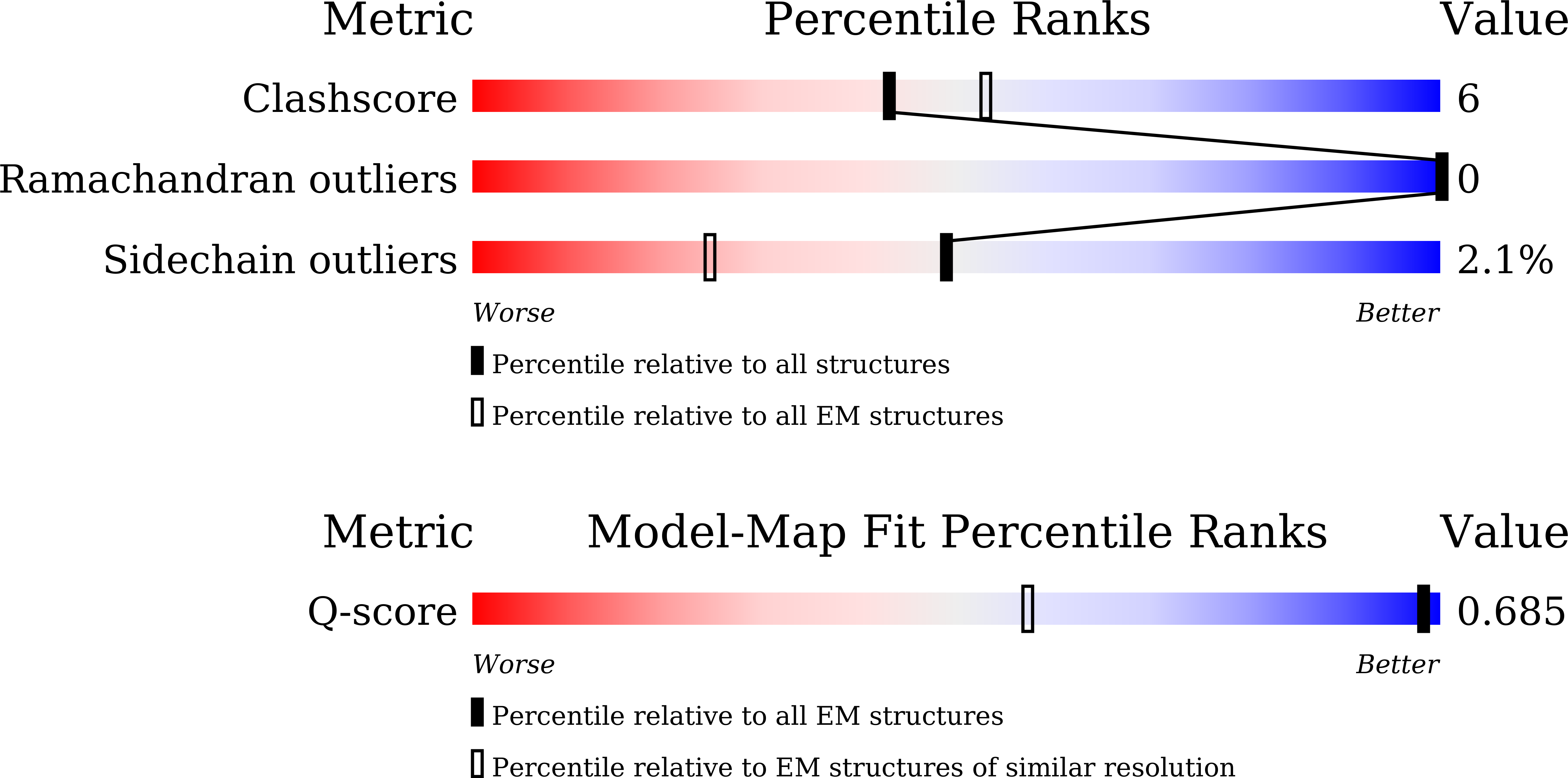
Cryo-EM structure of LH1-RC from Rhodovulum sulfidophilum
Biological Source:
Source Organism(s):
Method Details:
Experimental Method:
Resolution:
1.81 Å
Aggregation State:
PARTICLE
Reconstruction Method:
SINGLE PARTICLE
Macromolecular Entities
Structures with similar UniProt ID
Protein Blast
Polymer Type:polypeptide(L)
Molecule:Antenna pigment protein beta
Chain IDs:A (auth: 0), C (auth: 2), E (auth: 4), G (auth: 6), I (auth: 8), L (auth: B), O (auth: E), Q (auth: G), T (auth: J), X (auth: N), Z (auth: P), BA (auth: R), DA (auth: T), FA (auth: V), HA (auth: X), JA (auth: Z)
Chain Length:48
Number of Molecules:16
Biological Source:Rhodovulum sulfidophilum DSM 1374
Structures with similar UniProt ID
Protein Blast
Polymer Type:polypeptide(L)
Molecule:Antenna pigment protein alpha
Chain IDs:B (auth: 1), D (auth: 3), F (auth: 5), H (auth: 7), J (auth: 9), K (auth: A), N (auth: D), P (auth: F), S (auth: I), U (auth: K), Y (auth: O), AA (auth: Q), CA (auth: S), EA (auth: U), GA (auth: W), IA (auth: Y)
Chain Length:54
Number of Molecules:16
Biological Source:Rhodovulum sulfidophilum DSM 1374
Structures with similar UniProt ID
Protein Blast
Polymer Type:polypeptide(L)
Molecule:Photosynthetic reaction cente
Chain IDs:M (auth: C)
Chain Length:356
Number of Molecules:1
Biological Source:Rhodovulum sulfidophilum DSM 1374
Protein Blast
Polymer Type:polypeptide(L)
Molecule:photosynthetic reaction cente
Chain IDs:R (auth: H)
Chain Length:260
Number of Molecules:1
Biological Source:Rhodovulum sulfidophilum DSM 1374
Structures with similar UniProt ID
Protein Blast
Polymer Type:polypeptide(L)
Molecule:Reaction center protein L cha
Chain IDs:V (auth: L)
Chain Length:284
Number of Molecules:1
Biological Source:Rhodovulum sulfidophilum DSM 1374
Structures with similar UniProt ID
Protein Blast
Polymer Type:polypeptide(L)
Molecule:Reaction center protein M cha
Chain IDs:W (auth: M)
Chain Length:321
Number of Molecules:1
Biological Source:Rhodovulum sulfidophilum DSM 1374
Protein Blast
Polymer Type:polypeptide(L)
Molecule:NADH:ubiquinone oxidoreductas
Chain IDs:KA (auth: a)
Chain Length:54
Number of Molecules:1
Biological Source:Rhodovulum sulfidophilum DSM 1374
Modified Residue
Compound ID Chain ID Parent Comp ID Details 2D Image
FME D MET modified residue
Primary Citation
Structural insights into the photochemistry of the LH1-RC complex from the marine purple phototrophic bacterium Rhodovulum sulfidophilum.
Commun Biol 9 ? ? (2026)
PMID: 41772109 DOI: 10.1038/s42003-026-09755-z

Abstact

The marine purple nonsulfur phototrophic bacterium Rhodovulum (Rdv.) sulfidophilum (Alphaproteobacteria) has been a model organism for bacterial photosynthesis research because of its unusual ability to grow phototrophically (anoxic/light) using high concentrations of inorganic or organic sulfur compounds as electron donors or by respiration under fully oxic conditions. Here we present a 1.81 A-resolution cryo-EM structure of the light-harvesting 1-reaction center (LH1-RC) photocomplex from the Rdv. sulfidophilum type strain W4 with a focus on RC structure and function. The Rdv. sulfidophilum RC is characterized by its cytochrome (Cyt) subunit that contains three heme groups and is anchored by its intact N-terminal domain in the membrane. In contrast to a methionine as the 6th axial ligand to the heme-2 in other bacterial RC-bound triheme and tetraheme Cyt subunits, the outmost heme-2 in the Rdv. sulfidophilum Cyt subunit is ligated by a cysteine residue, resulting in a significant downshift of reduction potential of 470 mV compared to that of a methionine-ligated heme-2. A nonheme Fe ligated by a histidine of the Cyt subunit and five water molecules was identified in close proximity to heme-2, implying a potential role in electron transport from soluble electron donors to heme-2. The Rdv. sulfidophilum LH1 complex forms an open ring structure consisting of 16 alphabeta-subunits with a gap formed where the N-terminal transmembrane domain of the RC Cyt subunit and a newly identified protein with three helical domains (designated as protein-3h) are located. Protein-3h corresponds to the truncated N-terminal fragment of a gene product encoded by the pseudo-gene urf1 in the NADH:ubiquinone oxidoreductase (complex I) nuo operon in the genome of Rdv. sulfidophilum W4. Genes urf1 are also found in other purple nonsulfur bacteria and in aerobic anoxygenic phototrophic bacteria, and their putative products all share a common structural motif of N-terminal transmembrane U-shaped tandem helices. Based on structural and spectroscopic data, possible electron transfer pathways between the Rdv. sulfidophilum RC Cyt subunit and soluble electron donors and potential roles of protein-3h in the structural integrity of LH1-RC are discussed.

Legend

Protein

Chemical

Disease

Primary Citation of related structures
Feedback Form
Name
Email
Institute
Feedback