9W7A image
Deposition Date 2025-08-06
Release Date 2026-04-01
Last Version Date 2026-04-01
Entry Detail
PDB ID:
9W7A
Keywords:
Title:
Crystal structure of L-galactose dehydrogenase from Luteolibacter sp. strain LG18 in complex with L-glucose and NADP+
Biological Source:
Source Organism(s):
Expression System(s):
Method Details:
Experimental Method:
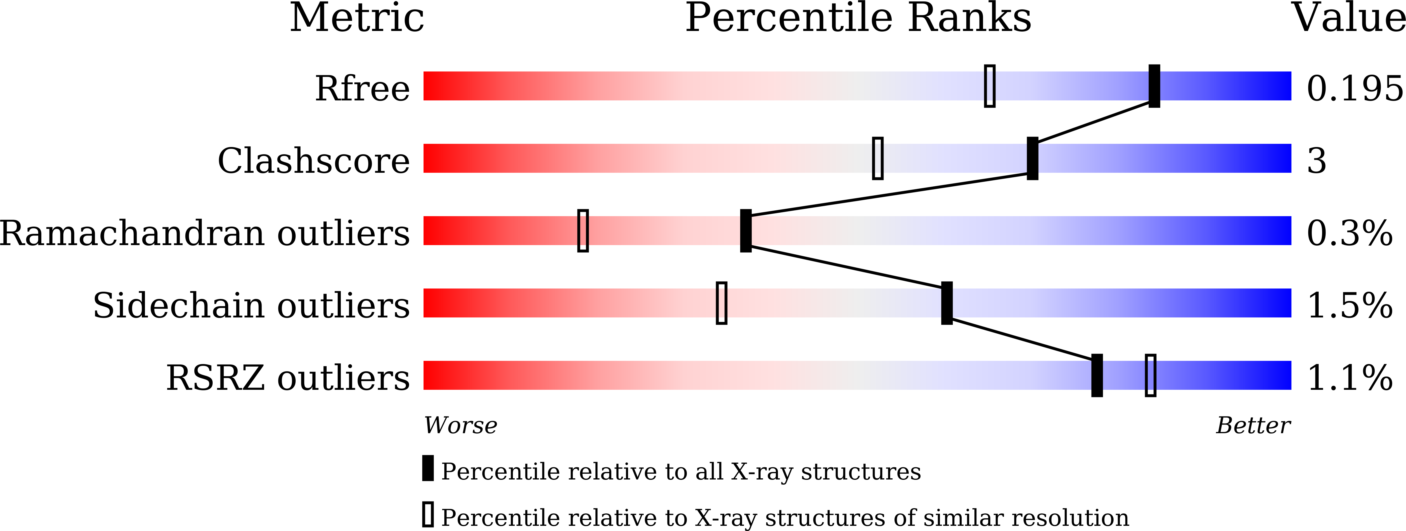
Resolution:
1.56 Å
R-Value Free:
0.18
R-Value Work:
0.15
Space Group:
P 21 21 21
Macromolecular Entities
Protein Blast
Polymer Type:polypeptide(L)
Molecule:L-galactose dehydrogenase
Chain IDs:A, B
Chain Length:349
Number of Molecules:2
Biological Source:Luteolibacter sp. LG18
Primary Citation
Characterization of bacterial l-galactose dehydrogenase with l-glucose dehydrogenase activity from Luteolibacter sp. strain LG18.
Biosci. Biotechnol. Biochem. ? ? ? (2026)
PMID: 41854348 DOI: 10.1093/bbb/zbag041

Abstact

The crystal structure of l-galactose dehydrogenase (LGDH) with l-glucose dehydrogenase activity from Luteolibacter sp. strain LG18 (Lu-LGDH) was determined in complex with l-galactose or l-glucose and NADP(+). This structural analysis identified key residues involved in substrate binding, and alanine-substituted mutants demonstrated the roles of these residues, including Tyr56, acting as potential general base within the catalytic tetrad. Unlike plant enzymes that show a preference for NAD(+), Lu-LGDH exhibits a marked preference for NADP(+) as a cofactor. This preference was attributed to the interaction of the phosphate group with Arg28, Thr269, and Asn274. The binding mode of l-glucose was similar to that of l-galactose. The C4 hydroxyl group (the structural difference between these pyranoses) was not used for substrate binding, which explains the dual activity of the enzyme. Furthermore, among the substrate-binding residues that were mutated, Arg308, which is not conserved among LGDHs, was crucial for the enzymatic activity.

Legend

Protein

Chemical

Disease

Primary Citation of related structures
Feedback Form
Name
Email
Institute
Feedback