9UPD image
Deposition Date 2025-04-28
Release Date 2025-07-16
Last Version Date 2025-07-16
Entry Detail
PDB ID:
9UPD
Title:
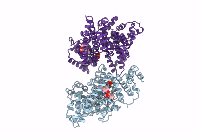
Crystal structure of human serum albumin complex with nateglinide
Biological Source:
Source Organism(s):
Homo sapiens (Taxon ID: 9606)
Expression System(s):
Method Details:
Experimental Method:
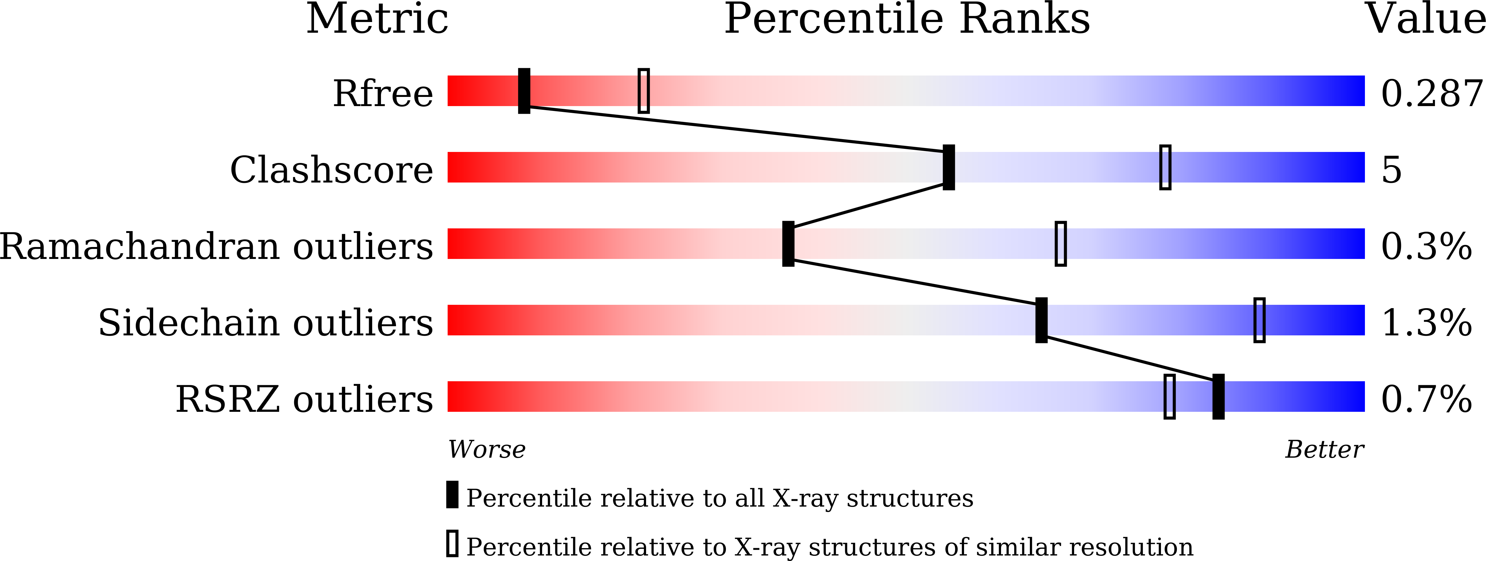
Resolution:
2.80 Å
R-Value Free:
0.28
R-Value Work:
0.24
R-Value Observed:
0.24
Space Group:
P 1 21 1
Macromolecular Entities
Structures with similar UniProt ID
Protein Blast
Polymer Type:polypeptide(L)
Molecule:Serum albumin
Gene (Uniprot):ALB
Chain IDs:A, B
Chain Length:585
Number of Molecules:2
Biological Source:Homo sapiens
Primary Citation
Insights into Dual Binding Modes of Nateglinide to Human Serum Albumin.
Acs Med.Chem.Lett. 16 1619 1625 (2025)
PMID: 40832515 DOI: 10.1021/acsmedchemlett.5c00277

Abstact

Nateglinide is a short-acting insulin secretagogue clinically used for the treatment of type 2 diabetes mellitus. Nateglinide exhibits a high plasma protein binding rate of approximately 98%, primarily binding to subdomain IIIA of human serum albumin (HSA). Here, we determined the crystal structure of the HSA-nateglinide complex at 2.80 Å resolution, revealing dual binding modes of nateglinide within the same binding site. To evaluate the stability of these alternative binding modes, molecular dynamics simulations were conducted, and binding free energies were calculated using the molecular mechanics Poisson-Boltzmann surface area method. The calculated binding free energy values were consistent with experimentally determined data. Our results highlight the structural adaptability of HSA and emphasize the possibility of alternative ligand binding modes existing within a single binding site, as well as the importance of identifying and characterizing these modes to enhance our understanding of drug-plasma protein interactions.

Legend

Protein

Chemical

Disease

Primary Citation of related structures
Feedback Form
Name
Email
Institute
Feedback