Deposition Date
2025-06-04
Release Date
2026-05-06
Last Version Date
2026-05-06
Entry Detail
PDB ID:
9REZ
Keywords:
Title:
Structure of PcuC from Cereibacter sphaeroides in its apo form
Biological Source:
Source Organism(s):
Cereibacter sphaeroides (Taxon ID: 1063)
Expression System(s):
Method Details:
Experimental Method:
Resolution:
1.05 Å
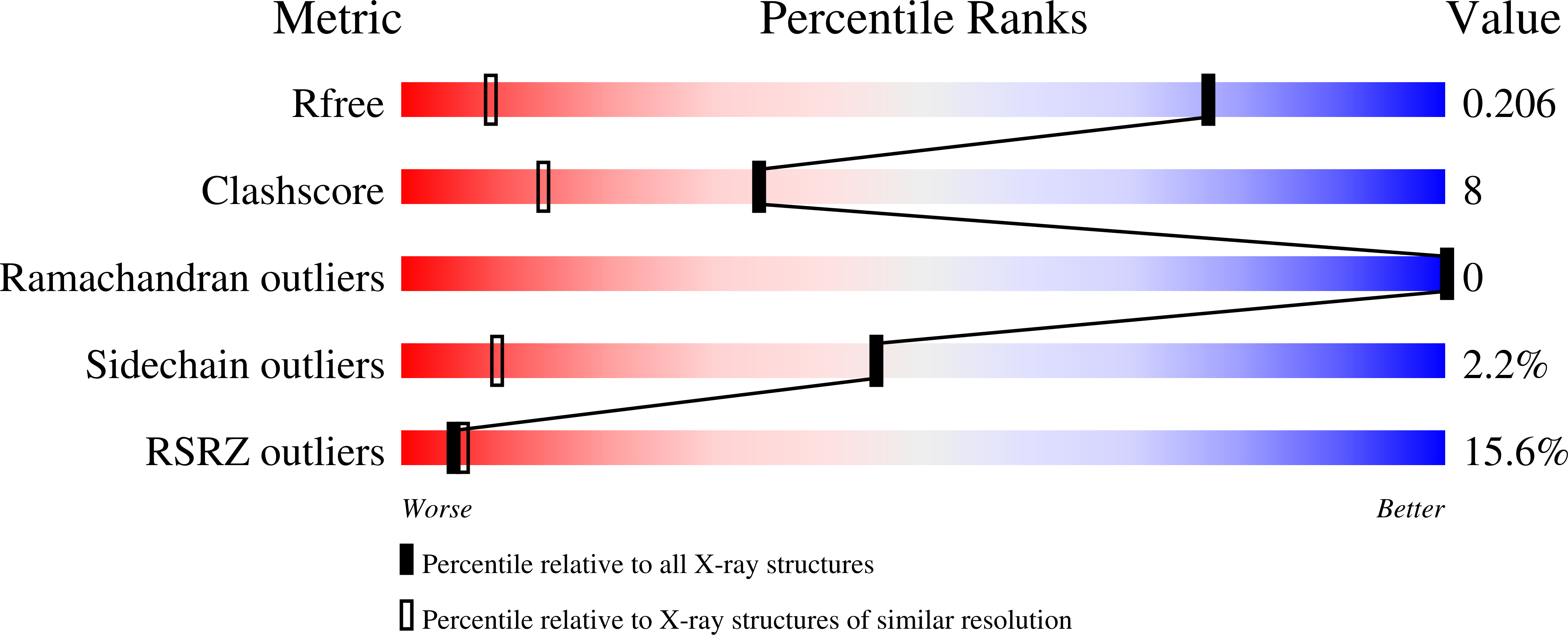
R-Value Free:
0.20
R-Value Work:
0.18
Space Group:
P 43 21 2