9PX9 image
Deposition Date 2025-08-05
Release Date 2026-05-13
Last Version Date 2026-05-13
Entry Detail
PDB ID:
9PX9
Keywords:
Title:
ATTR V122I cardiac amyloid fibrils
Biological Source:
Source Organism(s):
Homo sapiens (Taxon ID: 9606)
Method Details:
Experimental Method:
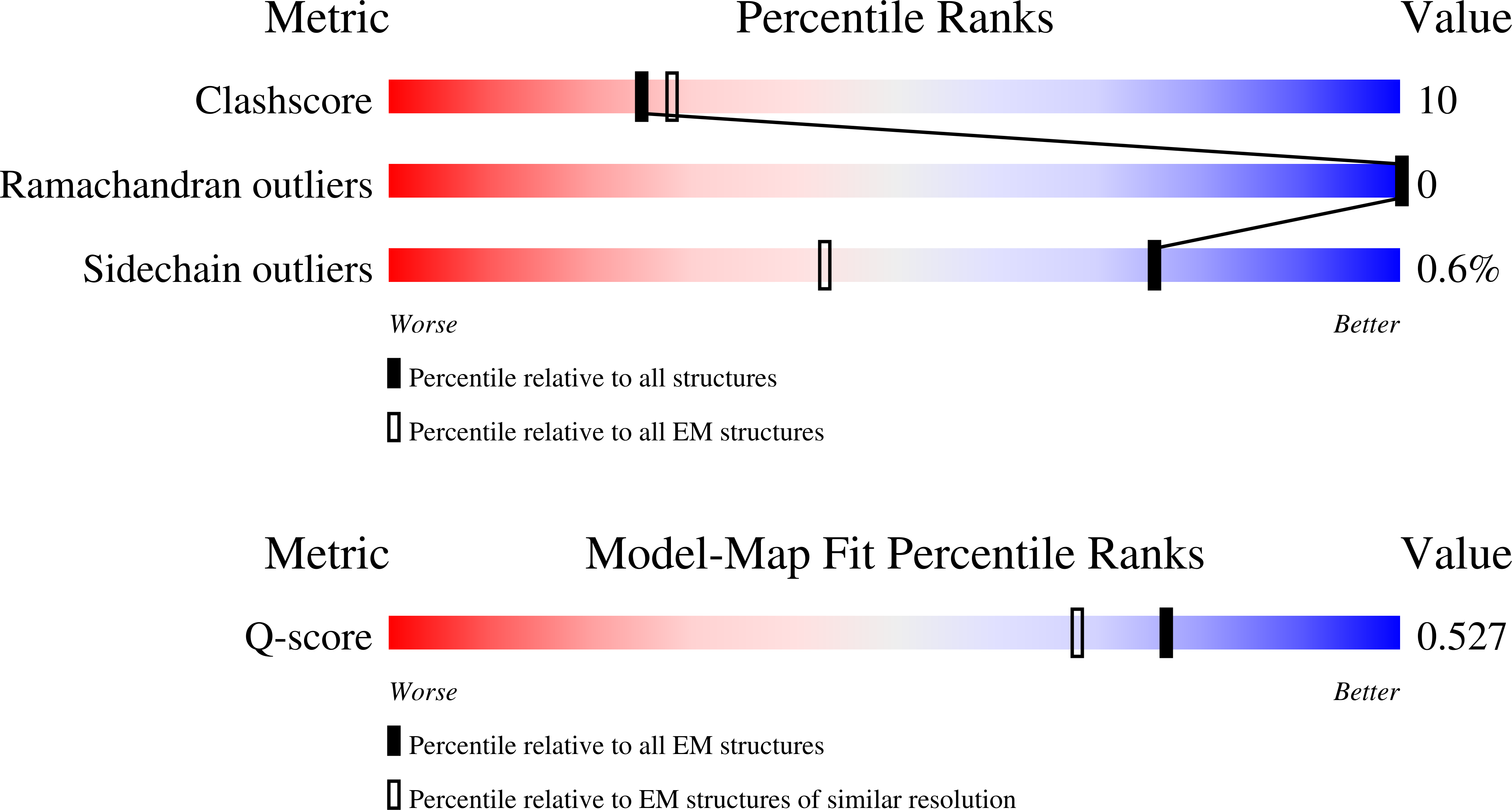
Resolution:
3.00 Å
Aggregation State:
FILAMENT
Reconstruction Method:
HELICAL
Macromolecular Entities
Structures with similar UniProt ID
Protein Blast
Polymer Type:polypeptide(L)
Molecule:Transthyretin
Gene (Uniprot):TTR
Chain IDs:A, B, C, D, E, F
Chain Length:147
Number of Molecules:6
Biological Source:Homo sapiens
Ligand Molecules
Primary Citation
Three-Dimensional Visualization and Proteomic Analysis of Human Cardiac Transthyretin Amyloidosis Tissue Reveals Microangiopathy and Capillary Occlusion.
J Am Heart Assoc ? e042248 e042248 (2026)
PMID: 42089154 DOI: 10.1161/JAHA.125.042248

Abstact

BACKGROUND: Transthyretin amyloidosis (ATTR) is a degenerative disease affecting the heart and other organs. Transthyretin (TTR) aggregation is a driver of ATTR pathology, but the mechanism is poorly understood. We used proteomics and tissue clearing technology on wild-type (WT) human cardiac (WT/WT) and V122I human cardiac (V122I/WT) tissue to better understand TTR cardiomyopathy. METHODS: Flash-frozen cardiac tissue slices from human subjects with end-stage WT-TTR cardiomyopathy, end-stage V122I TTR cardiomyopathy, and an age-matched control were used. Fibrils and tissue proteomes were extracted and assessed by bottom-up proteomics. Tissue clearing was performed using a lauryl sulfate-based lipid removal strategy. Slices were stained using indirect immunofluorescence against targets identified by proteomics. TTR deposits were imaged by antibody and AmyTracker 480 staining. Structures of ATTR fibrils were characterized using cryogenic electron microscopy. RESULTS: Proteomic analysis revealed high abundance of TTR, proteins associated with amyloid fibrils, as well as angiogenic, hemostatic, and complement cascade-associated proteins. Three-dimensional imaging revealed loss of normal microvascular architecture, regions of hypervascularization and hypovascularization, and microvascular obstruction by capillary thrombosis. ATTR fibrils adopted the spearhead fold and were decorated with collagen VI, an extracellular matrix component. CONCLUSIONS: Based on our imaging and proteomic data, we hypothesize that ATTR cardiomyopathy is a microangiopathy driven by capillary bed thromboinflammation and dysregulated angiogenic revascularization. In this model, increased capillary permeability exposes components of the vascular basement membrane to misfolded TTR. These components promote aggregation and stabilize amyloid fibrils. Congestion of the vascular basement membrane prevents appropriate revascularization, reducing cardiac exertional capacity over time, leading to heart failure.

Legend

Protein

Chemical

Disease

Primary Citation of related structures
Feedback Form
Name
Email
Institute
Feedback