Deposition Date
2024-10-28
Release Date
2025-06-11
Last Version Date
2025-06-11
Entry Detail
PDB ID:
9H80
EMDB ID:
Keywords:
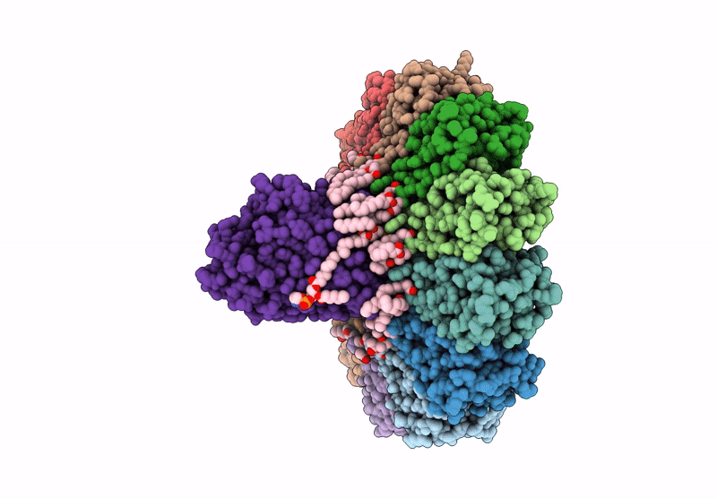
Title:
Structure of the outer membrane exopolysaccharide transporter PelBC
Biological Source:
Source Organism(s):
Pseudomonas aeruginosa (Taxon ID: 287)
Expression System(s):
Method Details:
Experimental Method:
Resolution:
2.50 Å
Aggregation State:
PARTICLE
Reconstruction Method:
SINGLE PARTICLE