9FVK image
Deposition Date 2024-06-27
Release Date 2025-07-09
Last Version Date 2026-01-28
Entry Detail
PDB ID:
9FVK
Keywords:
Title:
Crystal structure of amyloidogenic light chain AL-55 in open conformation.
Biological Source:
Source Organism(s):
Homo sapiens (Taxon ID: 9606)
Expression System(s):
Method Details:
Experimental Method:
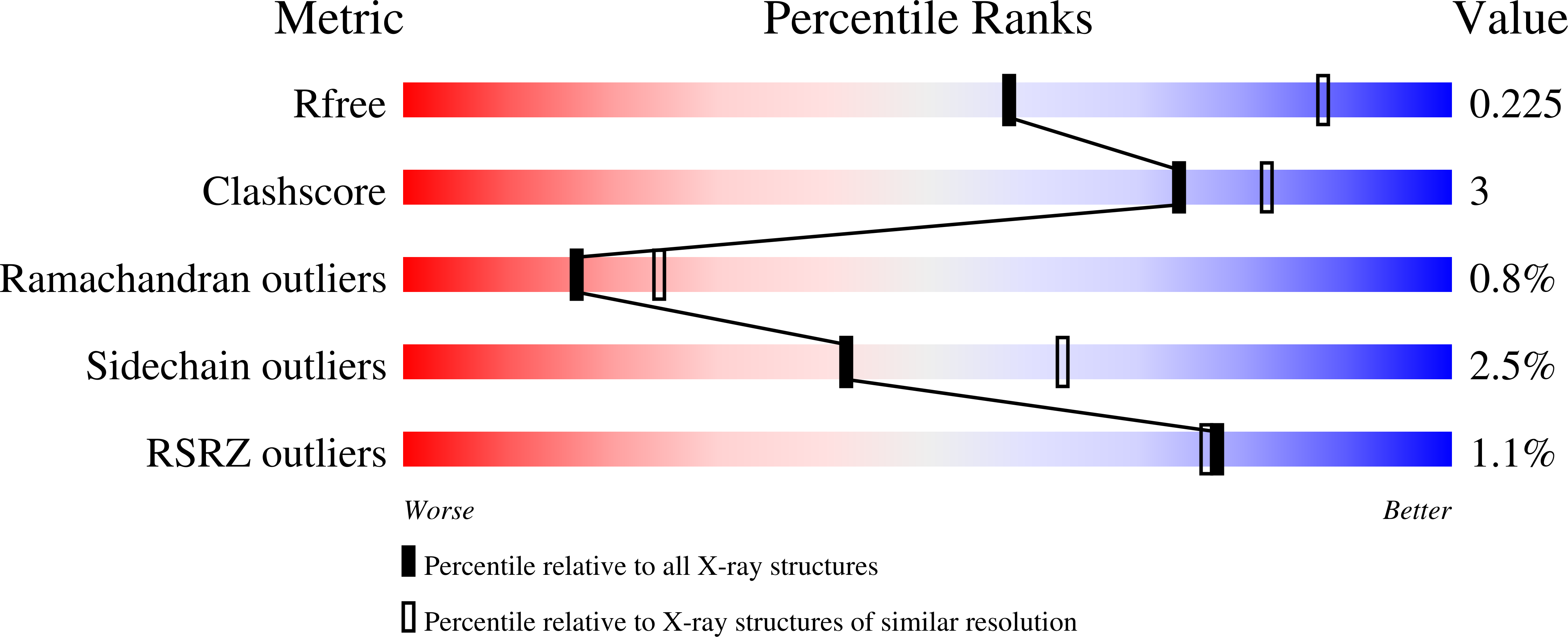
Resolution:
2.64 Å
R-Value Free:
0.22
R-Value Work:
0.19
R-Value Observed:
0.19
Space Group:
C 2 2 21
Macromolecular Entities
Structures with similar UniProt ID
Protein Blast
Polymer Type:polypeptide(L)
Molecule:AL55
Chain IDs:A, B, C
Chain Length:217
Number of Molecules:3
Biological Source:Homo sapiens
Primary Citation
The Critical Role of the Variable Domain in Driving Proteotoxicity and Aggregation in Full-length Light Chains.
J. Mol. Biol. 437 168958 168958 (2025)
PMID: 39842712 DOI: 10.1016/j.jmb.2025.168958

Abstact

Light chain (AL) amyloidosis is the most common systemic amyloid disease characterized by abnormal accumulation of amyloid fibrils derived from immunoglobulin light chains (LCs). Both full-length (FL) LCs and their isolated variable (VL) and constant (CL) domains contribute to amyloid deposits in multiple organs, with the VL domain predominantly forming the fibril core. However, the role and interplay of these domains in amyloid aggregation and toxicity are poorly understood. Characterizing the amyloidogenic λ6-LC AL55, this study explores the properties of both FL and isolated domains and compares them with the available patient-derived data. FL AL55 biophysical features result from the interplay between its VL and CL domains where the limited VL-CL interface might play a major role. Slow refolding kinetic of FL confirms the unfolded VL domain as a kinetic trap possibly shifting the process towards misfolding. The X-ray structure of FL AL55 shows that VL domains may detach from the native dimeric assembly and establish non-native interdimeric interfaces. Additionally, isolated VL domains display significantly lower soluble toxicity compared to FL and do not form fibrils similar to those found ex vivo. Thus the data obtained in this work allowed us to draw a molecular sketch of the aggregation pathway for amyloidogenic LCs.

Legend

Protein

Chemical

Disease

Primary Citation of related structures
Feedback Form
Name
Email
Institute
Feedback