9D8S image
Deposition Date 2024-08-20
Release Date 2024-09-04
Last Version Date 2026-05-13
Entry Detail
PDB ID:
9D8S
Keywords:
Title:
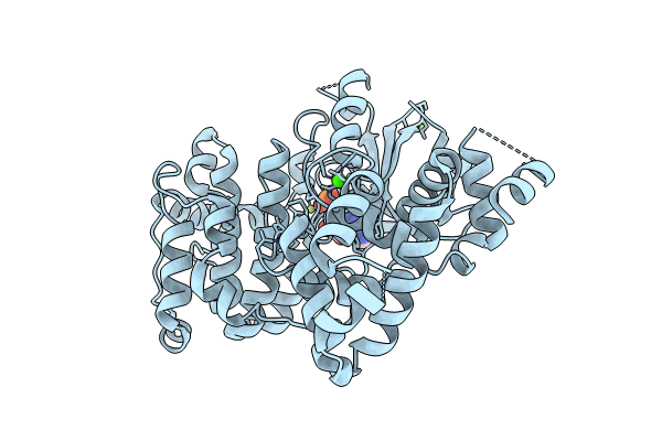
Crystal Structure of calcium-dependent protein kinase 1 (CDPK1) from Cryptosporidium parvum (AMP/Mg bound)
Biological Source:
Source Organism(s):
Expression System(s):
Method Details:
Experimental Method:
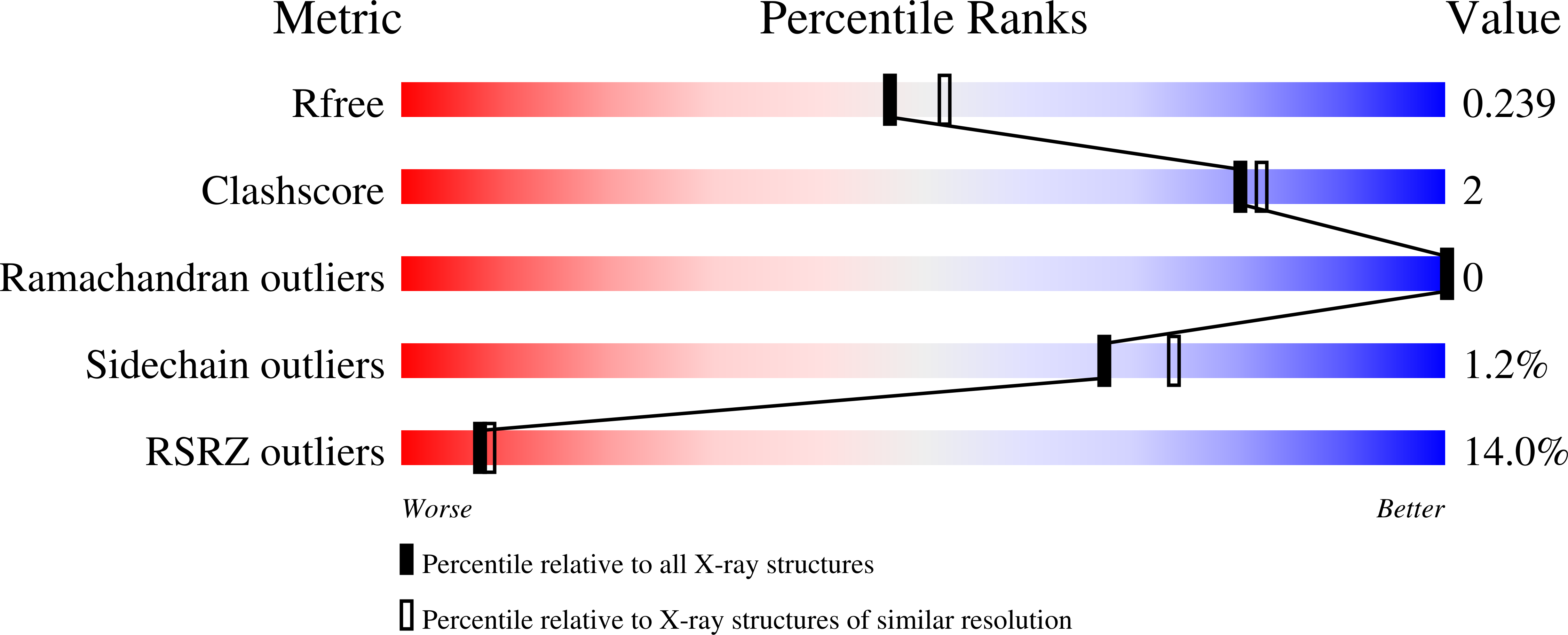
Resolution:
2.12 Å
R-Value Free:
0.24
R-Value Work:
0.20
R-Value Observed:
0.21
Space Group:
P 1 21 1
Macromolecular Entities
Structures with similar UniProt ID
Protein Blast
Polymer Type:polypeptide(L)
Molecule:Calmodulin-domain protein kin
Gene (Uniprot):cgd3_920
Chain IDs:A
Chain Length:492
Number of Molecules:1
Biological Source:Cryptosporidium parvum Iowa II
Primary Citation
Structure-guided design of calcium-dependent protein kinase 1 (CDPK1) inhibitors for cryptosporidiosis.
J.Infect.Dis. ? ? ? (2026)
PMID: 42081373 DOI: 10.1093/infdis/jiag242

Abstact

BACKGROUND: Calcium-dependent protein kinase 1 (CDPK1) has emerged as a protozoan specific target for the treatment of cryptosporidiosis. A previous study identified pyridopyrimidinones as new Cryptosporidium parvum (Cp) CDPK1 inhibitors with potent growth inhibition against C. parvum and C. hominis. Docking analyses suggested the unique positioning of the kinase's alphaC-helix could present refinement opportunities. METHODS: Compounds designed to optimize the pyridopyrimidinones focused on the back-pocket region predicted to be proximal to the alphaC-helix, the solvent exposed region and the ATP ribose-binding site. Designed derivatives were synthesized and assessed for CpCDPK1 and Src kinase inhibition and for Cryptosporidium spp., growth inhibition in mammalian cells. AMP/Mg+2 and three inhibitors were co-crystalized with CpCDPK1, and two inhibitors were profiled for kinase selectivity. RESULTS: WIN 4-88 was identified with CpCDPK1 (IC50 = 0.056 muM), and growth inhibition of zoonotic C. parvum (NLuc EC50 = 0.042 muM), anthroponotic C. parvum (Tu114 EC50 = 0.030 muM), and C. hominis Tu502 (EC50 = 0.062 muM), as well as enhanced kinome selectivity. The crystal structures confirmed the predicted binding mode, indicating key interactions with hinge residue Y155, similar orientations of the solvent expose moieties, occupancy of the back-pocket near the alphaC-helix and for one inhibitor containing a solubilizing hydroxyethyl attached to the central heterocycle extension into the ATP ribose-binding site. CONCLUSIONS: The expanded structure-activity relationship and structural insights will potentially be applicable to other chemotypes with similar binding modes and will enhance development of CpCDPK1 inhibitors for the treatment of cryptosporidiosis.

Legend

Protein

Chemical

Disease

Primary Citation of related structures
Feedback Form
Name
Email
Institute
Feedback