

7ZIT image
Deposition Date 2022-04-08
Release Date 2022-10-26
Last Version Date 2024-10-23
Entry Detail
PDB ID:
7ZIT
Keywords:
Title:
14-3-3 in complex with SARS-COV2 N phospho-peptide
Biological Source:
Expression System(s):
Method Details:
Experimental Method:
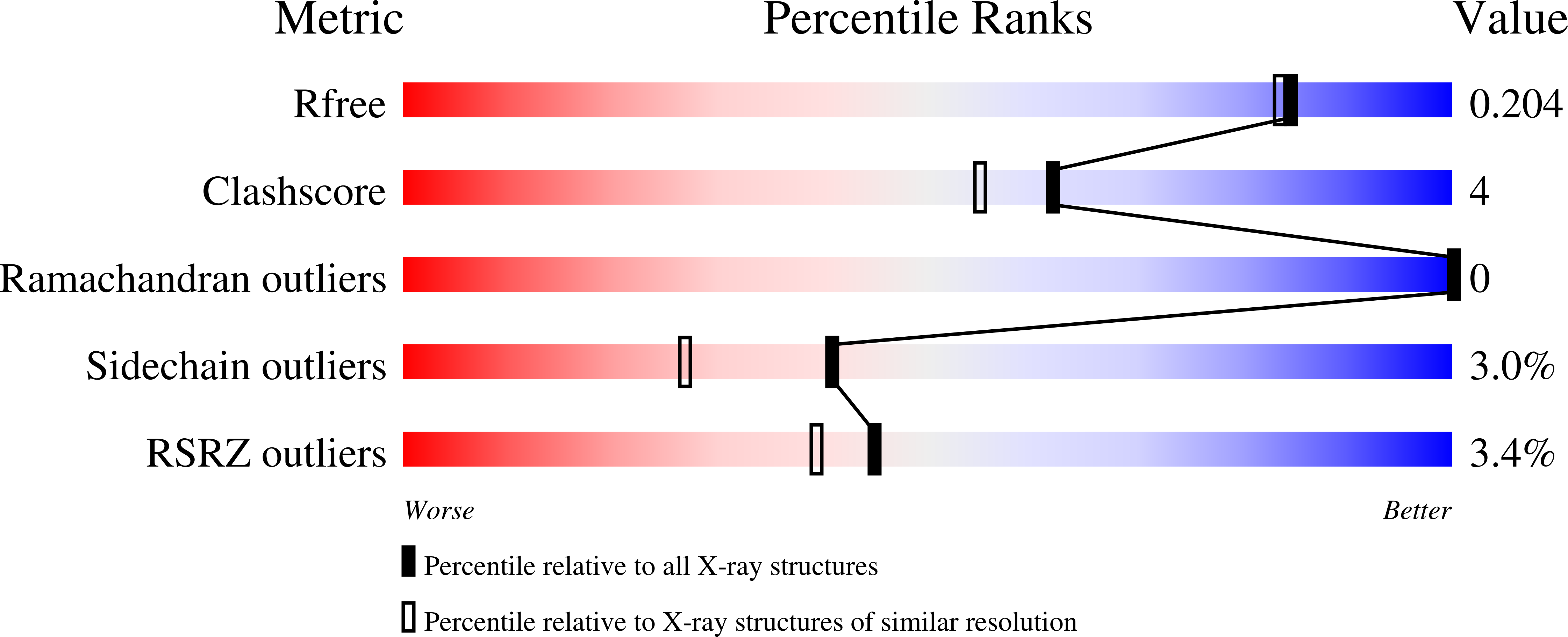
Resolution:
1.79 Å
R-Value Free:
0.20
R-Value Work:
0.18
R-Value Observed:
0.18
Space Group:
P 21 21 21
Macromolecular Entities
Structures with similar UniProt ID
Protein Blast
Polymer Type:polypeptide(L)
Molecule:14-3-3 protein zeta/delta
Gene (Uniprot):YWHAZ
Chain IDs:A, B
Chain Length:230
Number of Molecules:2
Biological Source:Homo sapiens
Structures with similar UniProt ID
Protein Blast
Polymer Type:polypeptide(L)
Molecule:Nucleoprotein
Gene (Uniprot):N
Chain IDs:C, D
Chain Length:7
Number of Molecules:2
Biological Source:Severe acute respiratory syndrome coronavirus 2
Modified Residue
Compound ID Chain ID Parent Comp ID Details 2D Image
SEP C SER modified residue
Primary Citation
Structural basis for SARS-CoV-2 nucleocapsid (N) protein recognition by 14-3-3 proteins.
J. Struct. Biol. 214 107879 107879 (2022)
PMID: 35781025 DOI: 10.1016/j.jsb.2022.107879

Abstact

14-3-3 proteins are important dimeric scaffolds that regulate the function of hundreds of proteins in a phosphorylation-dependent manner. The SARS-CoV-2 nucleocapsid (N) protein forms a complex with human 14-3-3 proteins upon phosphorylation, which has also been described for other coronaviruses. Here, we report a high-resolution crystal structure of 14-3-3 bound to an N phosphopeptide bearing the phosphoserine 197 in the middle. The structure revealed two copies of the N phosphopeptide bound, each in the central binding groove of each 14-3-3 monomer. A complex network of hydrogen bonds and water bridges between the peptide and 14-3-3 was observed explaining the high affinity of the N protein for 14-3-3 proteins.

Legend

Protein

Chemical

Disease

Primary Citation of related structures
Feedback Form
Name
Email
Institute
Feedback