7VEP image
Deposition Date 2021-09-09
Release Date 2022-09-14
Last Version Date 2026-03-18
Entry Detail
PDB ID:
7VEP
Keywords:
Title:
Crystal structure and biophysical characterization of TPR domain of EccA5 from ESX-5 pathway of Mycobacterium tuberculosis H37RVR
Biological Source:
Expression System(s):
Method Details:
Experimental Method:
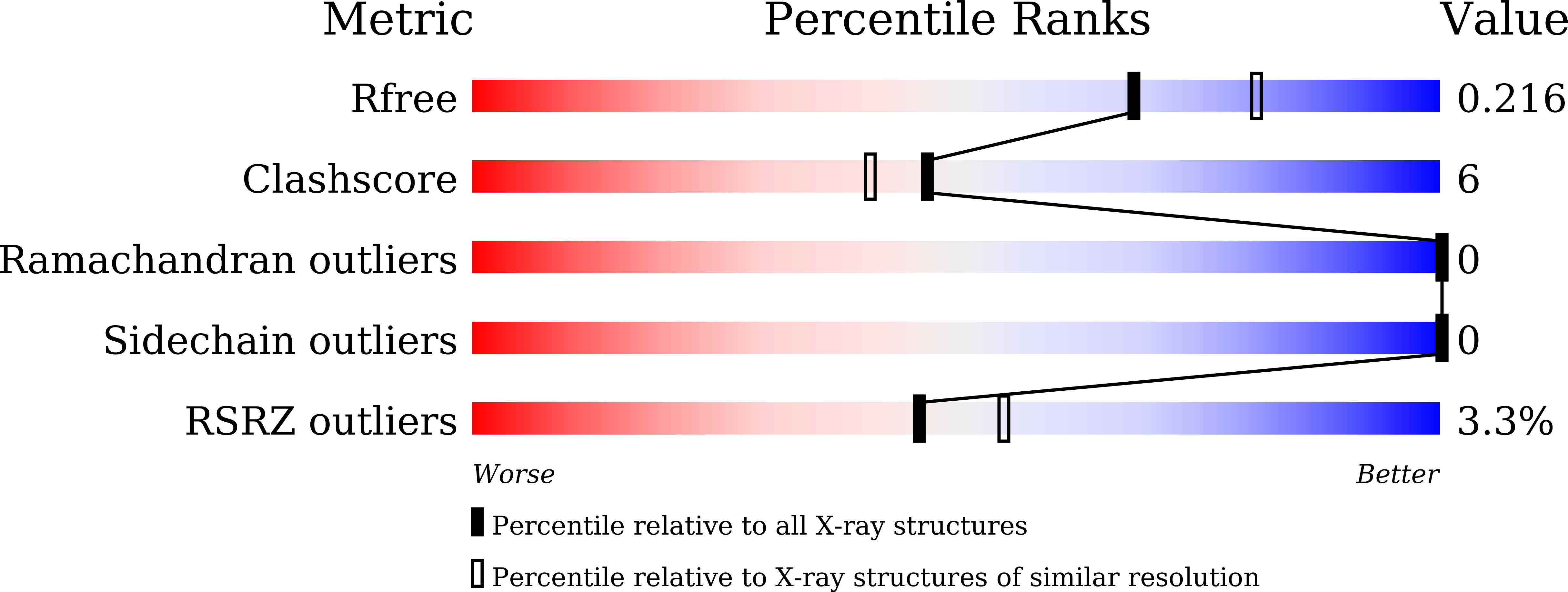
Resolution:
2.15 Å
R-Value Free:
0.21
R-Value Work:
0.18
R-Value Observed:
0.18
Space Group:
P 21 21 21
Macromolecular Entities
Structures with similar UniProt ID
Protein Blast
Polymer Type:polypeptide(L)
Molecule:ESX-5 secretion system protei
Gene (Uniprot):eccA5
Chain IDs:A
Chain Length:281
Number of Molecules:1
Biological Source:Mycobacterium tuberculosis (strain ATCC 25618 / H37Rv)
Primary Citation
The crystal structure of the TPR domain of the EccA5 ATPase and demonstration of its interaction with EspG5 from the mycobacterial ESX-5 pathway.
FEBS Lett. ? ? ? (2026)
PMID: 41761896 DOI: 10.1002/1873-3468.70315

Abstact

The ESX-5 secretion system in Mycobacterium tuberculosis exports PE/PPE virulence factors, with EccA5, an AAA+ ATPase, playing a pivotal role. We solved the crystal structure of EccA5's N-terminal TPR domain (EccA5NT) at 2.15 A, revealing a monomeric fold with six TPR motifs and a variable beta-finger. Biophysical studies, including SAXS and size exclusion chromatography, confirm its monomeric state. A flexible loop (residues 137-148) suggests dynamic substrate interactions. SPR, SAXS and in silico docking show moderate binding (K(D) = 3.43 mum) between EccA5NT's beta-finger and EspG5's beta2-beta3 loop, indicating a role in PE/PPE-EspG5 complex disassembly. These findings elucidate the role of EccA5 in ESX-5-mediated secretion.

Legend

Protein

Chemical

Disease

Primary Citation of related structures
Feedback Form
Name
Email
Institute
Feedback