7VA1 image
Deposition Date 2021-08-27
Release Date 2023-03-08
Last Version Date 2023-11-29
Entry Detail
PDB ID:
7VA1
Keywords:
Title:
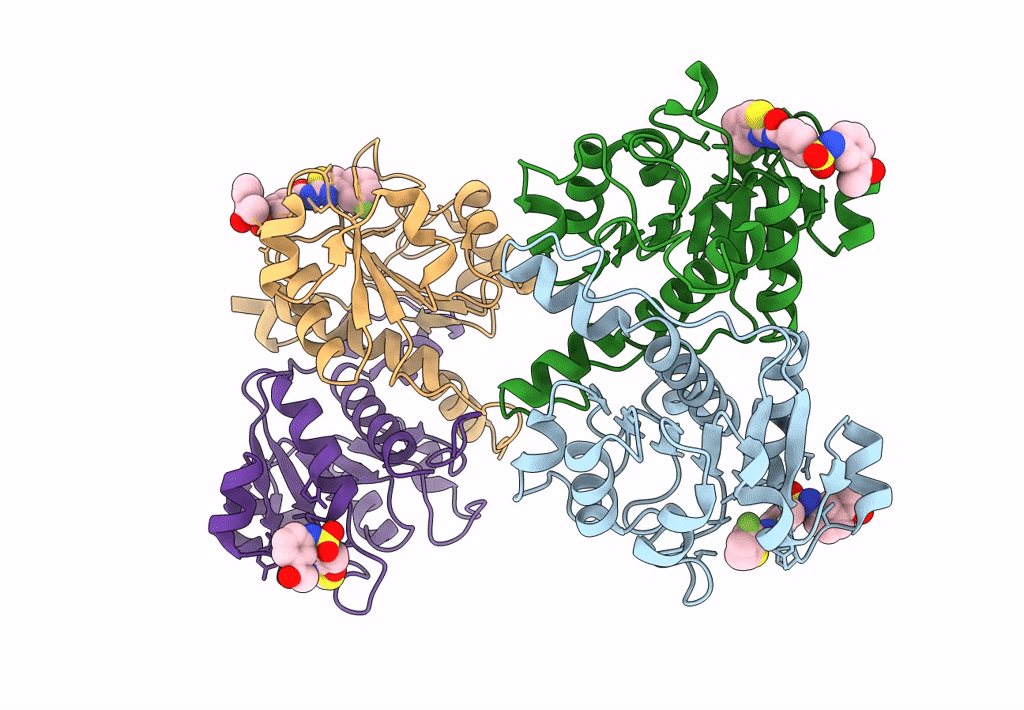
Crystal structure of human 3-phosphoglycerate dehydrogenase in complex with GDD-04-35
Biological Source:
Source Organism(s):
Homo sapiens (Taxon ID: 9606)
Expression System(s):
Method Details:
Experimental Method:
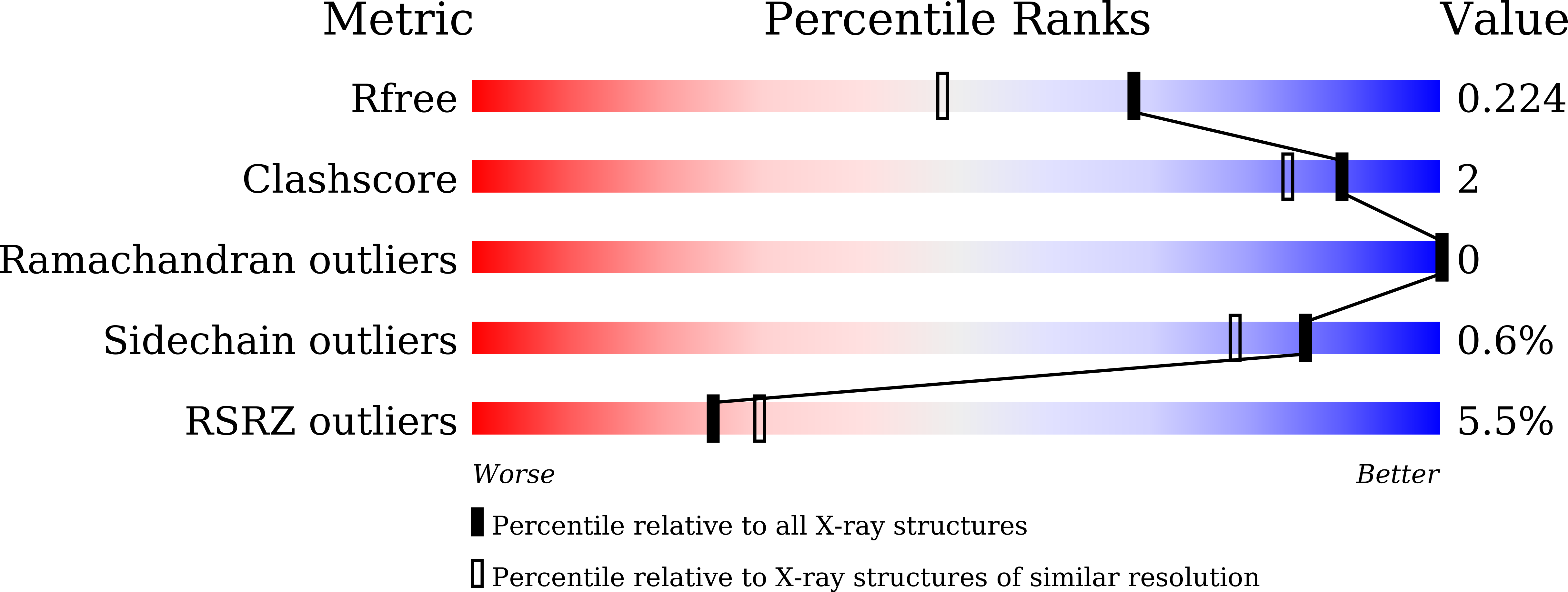
Resolution:
1.74 Å
R-Value Free:
0.22
R-Value Work:
0.19
R-Value Observed:
0.19
Space Group:
P 1 21 1
Macromolecular Entities
Structures with similar UniProt ID
Protein Blast
Polymer Type:polypeptide(L)
Molecule:D-3-phosphoglycerate dehydrog
Gene (Uniprot):PHGDH
Chain IDs:A (auth: B), B (auth: A), C, D
Chain Length:214
Number of Molecules:4
Biological Source:Homo sapiens
Ligand Molecules
Primary Citation
Discovery of Novel Drug-like PHGDH Inhibitors to Disrupt Serine Biosynthesis for Cancer Therapy.
J. Med. Chem. 66 285 305 (2023)
PMID: 36594670 DOI: 10.1021/acs.jmedchem.2c01202

Abstact

Being the rate-limiting enzyme within the serine biosynthesis pathway, phosphoglycerate dehydrogenase (PHGDH) is abnormally overexpressed in numerous malignant tumor cells and is a promising target for cancer treatment. Here, we report a series of novel PHGDH inhibitors using a focused compound screening and structural optimization approach. The lead compound D8 displayed good enzymatic inhibitory activity (IC50 = 2.8 ± 0.1 μM), high binding affinity (Kd = 2.33 μM), and sensitivity to the cell lines with the PHGDH gene amplification or overexpression. Furthermore, D8 was proven to restrict the de novo serine synthesis from glucose within MDA-MB-468 cells. X-ray crystallographic analysis, molecular dynamics simulations, and mutagenesis experiments on PHGDH revealed the binding site at D175 inside the NAD+-binding pocket. Finally, D8 exhibited excellent in vivo pharmacokinetic properties (F = 82.0%) and exerted evident antitumor efficacy in the PC9 xenograft mouse model.

Legend

Protein

Chemical

Disease

Primary Citation of related structures
Feedback Form
Name
Email
Institute
Feedback