Deposition Date
2022-01-21
Release Date
2023-08-16
Last Version Date
2026-03-04
Entry Detail
PDB ID:
7QVD
Keywords:
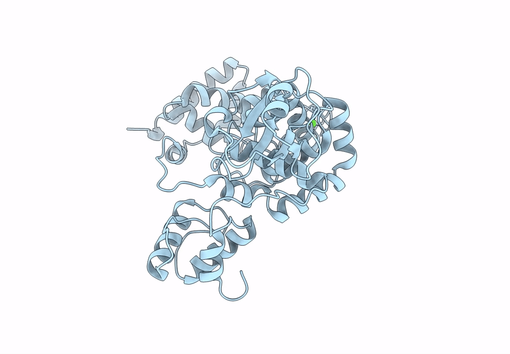
Title:
X-ray structure of the lytic transglycosylase SltB2 from Pseudomonas aeruginosa
Biological Source:
Source Organism(s):
Pseudomonas aeruginosa (Taxon ID: 287)
Expression System(s):
Method Details:
Experimental Method:
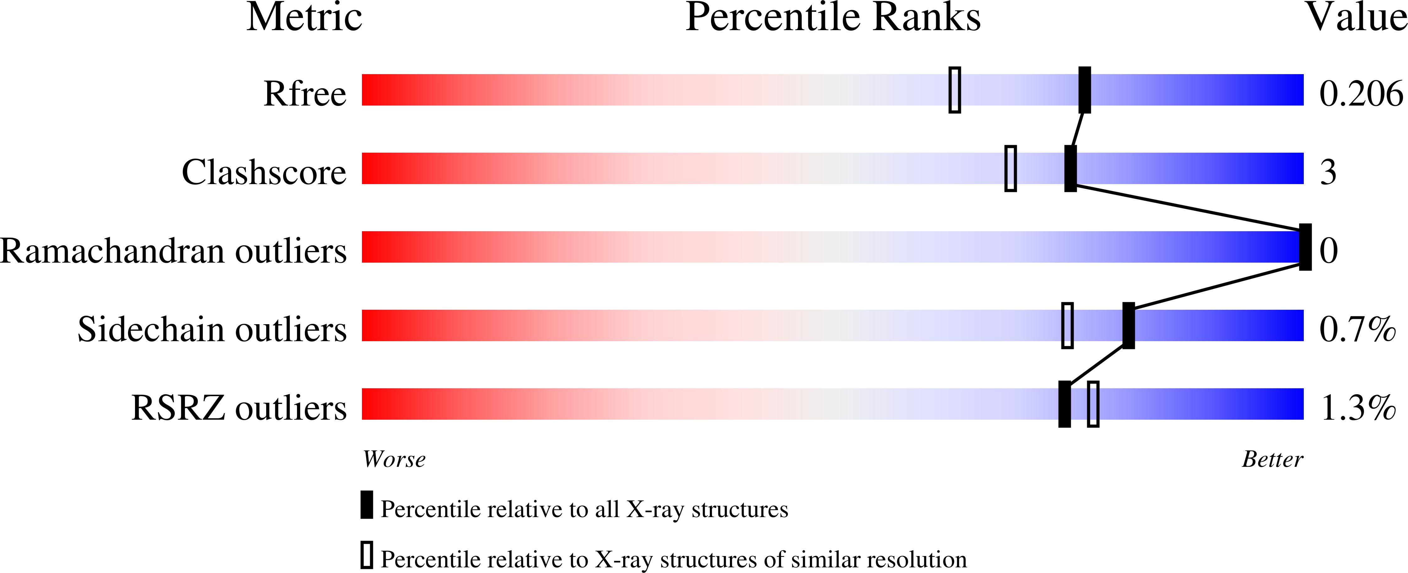
Resolution:
1.70 Å
R-Value Free:
0.19
R-Value Work:
0.16
Space Group:
P 1 21 1