7LVC image
Deposition Date 2021-02-24
Release Date 2021-03-17
Last Version Date 2026-02-11
Entry Detail
PDB ID:
7LVC
Keywords:
Title:
E. coli DHFR by Native Mn,P,S-SAD at Room Temperature
Biological Source:
Source Organism(s):
Expression System(s):
Method Details:
Experimental Method:
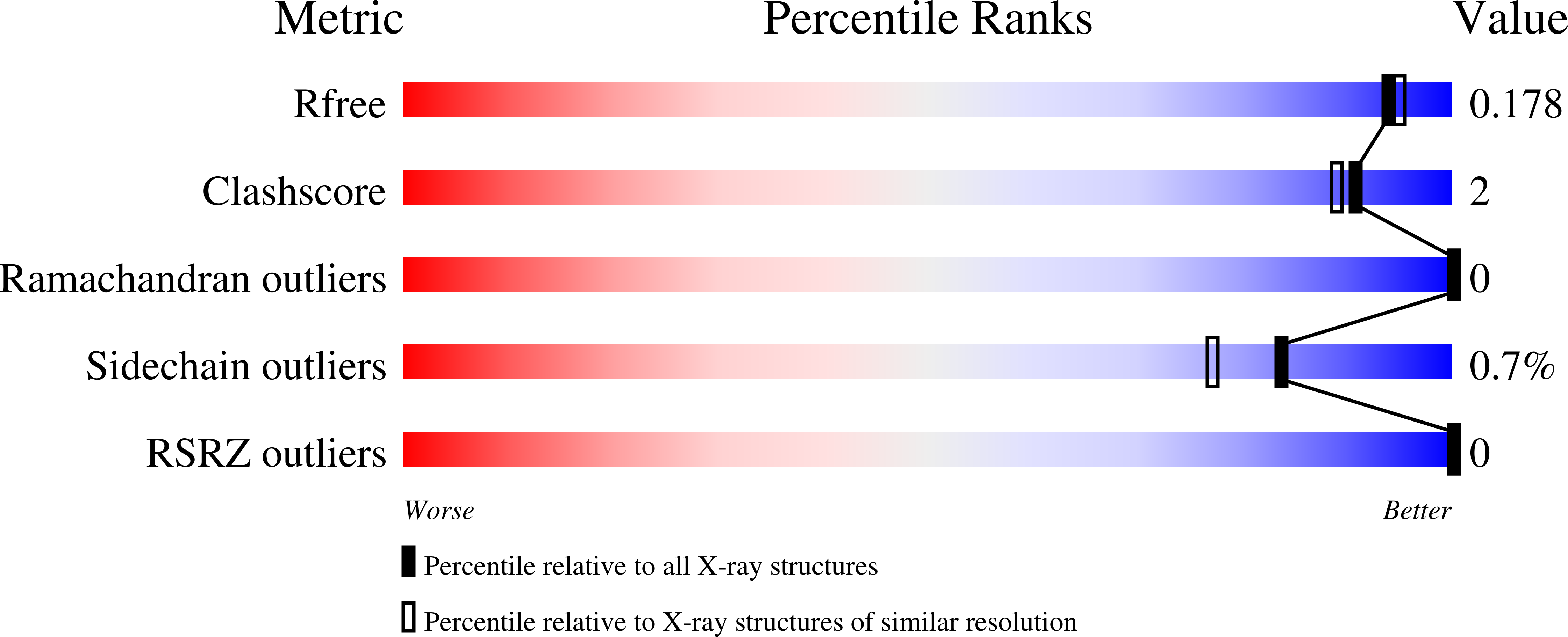
Resolution:
1.70 Å
R-Value Free:
0.17
R-Value Work:
0.13
R-Value Observed:
0.14
Space Group:
P 21 21 21
Macromolecular Entities
Structures with similar UniProt ID
Protein Blast
Polymer Type:polypeptide(L)
Molecule:Dihydrofolate reductase
Gene (Uniprot):folA
Chain IDs:A
Chain Length:159
Number of Molecules:1
Biological Source:Escherichia coli (strain K12)
Modified Residue
Compound ID Chain ID Parent Comp ID Details 2D Image
CSD A CYS modified residue
Primary Citation
Native SAD phasing at room temperature.
Acta Crystallogr D Struct Biol 78 986 996 (2022)
PMID: 35916223 DOI: 10.1107/S2059798322006799

Abstact

Single-wavelength anomalous diffraction (SAD) is a routine method for overcoming the phase problem when solving macromolecular structures. This technique requires the accurate measurement of intensities to determine differences between Bijvoet pairs. Although SAD experiments are commonly conducted at cryogenic temperatures to mitigate the effects of radiation damage, such temperatures can alter the conformational ensemble of the protein and may impede the merging of data from multiple crystals due to non-uniform freezing. Here, a strategy is presented to obtain high-quality data from room-temperature, single-crystal experiments. To illustrate the strengths of this approach, native SAD phasing at 6.55 keV was used to solve four structures of three model systems at 295 K. The resulting data sets allow automatic phasing and model building, and reveal alternate conformations that reflect the structure of proteins at room temperature.

Legend

Protein

Chemical

Disease

Primary Citation of related structures
Feedback Form
Name
Email
Institute
Feedback