7CT4 image
Deposition Date 2020-08-18
Release Date 2020-11-11
Last Version Date 2024-10-23
Entry Detail
PDB ID:
7CT4
Keywords:
Title:
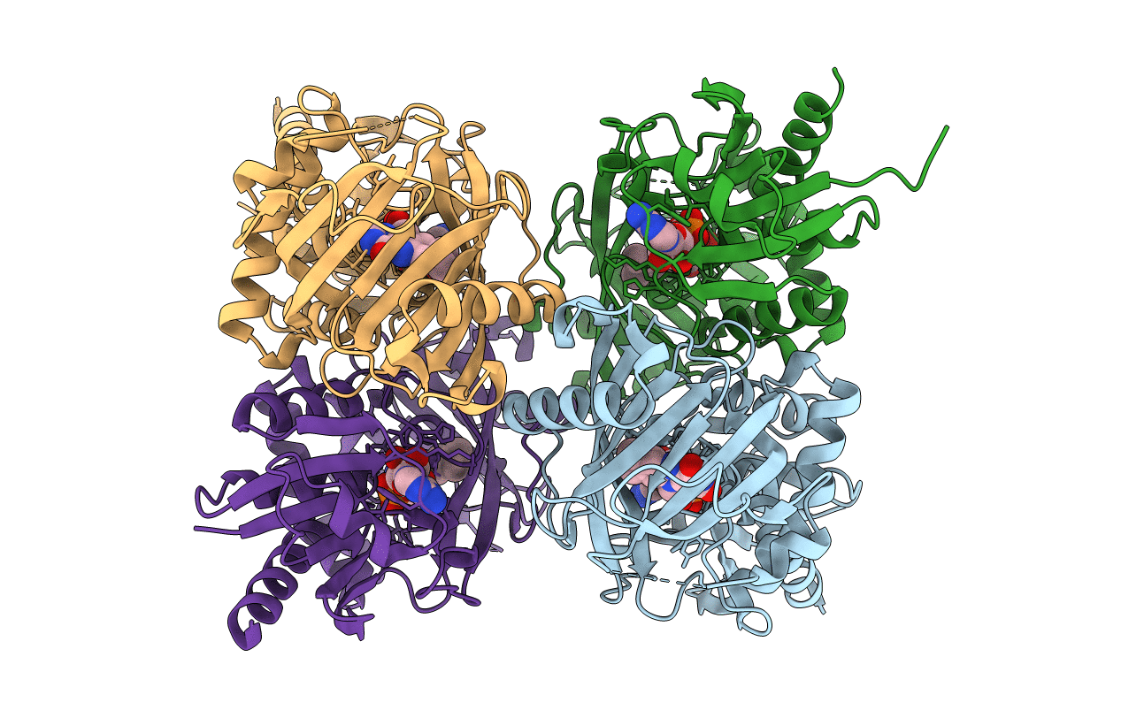
Crystal structure of D-amino acid oxidase from Rasamsonia emersonii strain YA
Biological Source:
Source Organism(s):
Expression System(s):
Method Details:
Experimental Method:
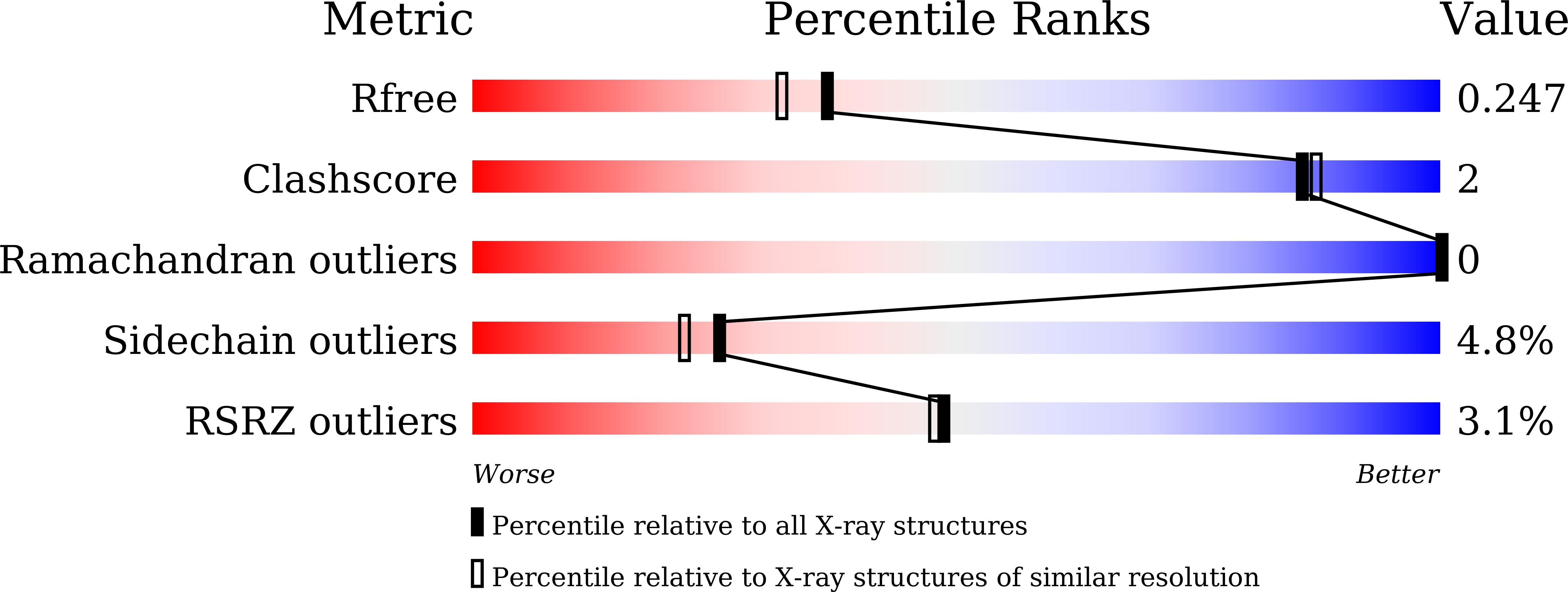
Resolution:
2.00 Å
R-Value Free:
0.24
R-Value Work:
0.20
R-Value Observed:
0.20
Space Group:
P 61
Macromolecular Entities
Structures with similar UniProt ID
Protein Blast
Polymer Type:polypeptide(L)
Molecule:D-amino acid oxidase
Gene (Uniprot):DAO1
Chain IDs:A, B, C, D
Chain Length:388
Number of Molecules:4
Biological Source:Talaromyces emersonii
Ligand Molecules
Primary Citation
X-ray structure analysis of a unique D-amino-acid oxidase from the thermophilic fungus Rasamsonia emersonii strain YA.
Acta Crystallogr. Sect. F Struct. Biol. Cryst. Commun. 76 517 523 (2020)
PMID: 33135670 DOI: 10.1107/S2053230X20013333

Abstact

D-Amino-acid oxidases (DAAOs) catalyze the oxidative deamination of neutral and basic D-amino acids. The DAAO from the thermophilic fungus Rasamsonia emersonii strain YA (ReDAAO) has a high thermal stability and a unique broad substrate specificity that includes the acidic D-amino acid D-Glu as well as various neutral and basic D-amino acids. In this study, ReDAAO was crystallized by the hanging-drop vapor-diffusion method and its crystal structure was determined at a resolution of 2.00 Å. The crystal structure of the enzyme revealed that unlike other DAAOs, ReDAAO forms a homotetramer and contains an intramolecular disulfide bond (Cys230-Cys285), suggesting that this disulfide bond is involved in the higher thermal stability of ReDAAO. Moreover, the structure of the active site and its vicinity in ReDAAO indicates that Arg97, Lys99, Lys114 and Ser231 are candidates for recognizing the side chain of D-Glu.

Legend

Protein

Chemical

Disease

Primary Citation of related structures
Feedback Form
Name
Email
Institute
Feedback