6MH9 image
Deposition Date 2018-09-17
Release Date 2019-11-13
Last Version Date 2023-10-11
Entry Detail
PDB ID:
6MH9
Keywords:
Title:
The crystal structure of the Staphylococcus aureus Fatty acid Kinase (Fak) B1 protein A121I mutant to 2.02 Angstrom resolution
Biological Source:
Source Organism(s):
Expression System(s):
Method Details:
Experimental Method:
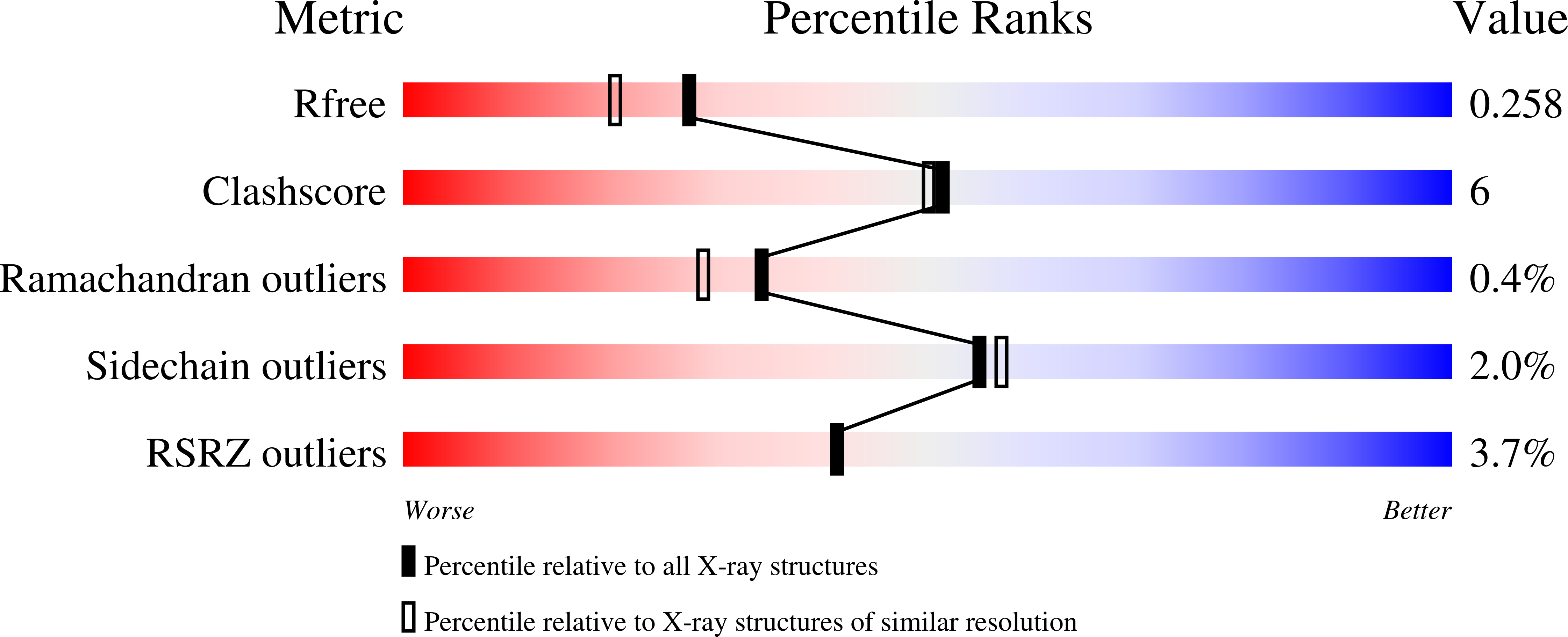
Resolution:
2.02 Å
R-Value Free:
0.25
R-Value Work:
0.20
R-Value Observed:
0.20
Space Group:
P 1
Macromolecular Entities
Structures with similar UniProt ID
Protein Blast
Polymer Type:polypeptide(L)
Molecule:Fatty Acid Kinase (Fak) B1 pr
Mutagens:A121I
Chain IDs:A, B
Chain Length:289
Number of Molecules:2
Biological Source:Staphylococcus aureus
Ligand Molecules
Primary Citation
Identification of structural transitions in bacterial fatty acid binding proteins that permit ligand entry and exit at membranes.
J. Biol. Chem. 298 101676 101676 (2022)
PMID: 35122790 DOI: 10.1016/j.jbc.2022.101676

Abstact

Fatty acid (FA) transfer proteins extract FA from membranes and sequester them to facilitate their movement through the cytosol. Detailed structural information is available for these soluble protein-FA complexes, but the structure of the protein conformation responsible for FA exchange at the membrane is unknown. Staphylococcus aureus FakB1 is a prototypical bacterial FA transfer protein that binds palmitate within a narrow, buried tunnel. Here, we define the conformational change from a "closed" FakB1 state to an "open" state that associates with the membrane and provides a path for entry and egress of the FA. Using NMR spectroscopy, we identified a conformationally flexible dynamic region in FakB1, and X-ray crystallography of FakB1 mutants captured the conformation of the open state. In addition, molecular dynamics simulations show that the new amphipathic α-helix formed in the open state inserts below the phosphate plane of the bilayer to create a diffusion channel for the hydrophobic FA tail to access the hydrocarbon core and place the carboxyl group at the phosphate layer. The membrane binding and catalytic properties of site-directed mutants were consistent with the proposed membrane docked structure predicted by our molecular dynamics simulations. Finally, the structure of the bilayer-associated conformation of FakB1 has local similarities with mammalian FA binding proteins and provides a conceptual framework for how these proteins interact with the membrane to create a diffusion channel from the FA location in the bilayer to the protein interior.

Legend

Protein

Chemical

Disease

Primary Citation of related structures
Feedback Form
Name
Email
Institute
Feedback